 Movie
Movie Controller

[English] 日本語
 Yorodumi
- PDB-1nap: THE CRYSTAL STRUCTURE OF RECOMBINANT HUMAN NEUTROPHIL-ACTIVATING ... -
+ Open data

- Basic information
| Entry | Database: PDB / ID: 1nap | ||||||
|---|---|---|---|---|---|---|---|
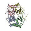
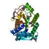
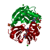
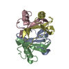
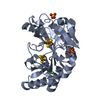
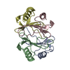
| Title | THE CRYSTAL STRUCTURE OF RECOMBINANT HUMAN NEUTROPHIL-ACTIVATING PEPTIDE-2 (M6L) AT 1.9-ANGSTROMS RESOLUTION | ||||||
 Components Components | NEUTROPHIL ACTIVATING PEPTIDE-2 | ||||||
 Keywords Keywords | CYTOKINE | ||||||
| Function / homology |  Function and homology information Function and homology informationD-glucose transmembrane transporter activity / D-glucose transmembrane transport / CXCR chemokine receptor binding / chemokine activity / Chemokine receptors bind chemokines / positive regulation of cell division / neutrophil chemotaxis / platelet alpha granule lumen / growth factor activity / tertiary granule lumen ...D-glucose transmembrane transporter activity / D-glucose transmembrane transport / CXCR chemokine receptor binding / chemokine activity / Chemokine receptors bind chemokines / positive regulation of cell division / neutrophil chemotaxis / platelet alpha granule lumen / growth factor activity / tertiary granule lumen / Platelet degranulation / antimicrobial humoral immune response mediated by antimicrobial peptide / cellular response to lipopolysaccharide / G alpha (i) signalling events / defense response to bacterium / inflammatory response / Neutrophil degranulation / : / extracellular region Similarity search - Function | ||||||
| Biological species |  Homo sapiens (human) Homo sapiens (human) | ||||||
| Method |  X-RAY DIFFRACTION / Resolution: 1.9 Å X-RAY DIFFRACTION / Resolution: 1.9 Å | ||||||
 Authors Authors | Malkowski, M.G. / Edwards, B.F.P. | ||||||
 Citation Citation | Journal: J.Biol.Chem. / Year: 1995 Title: The crystal structure of recombinant human neutrophil-activating peptide-2 (M6L) at 1.9-A resolution. Authors: Malkowski, M.G. / Wu, J.Y. / Lazar, J.B. / Johnson, P.H. / Edwards, B.F. #1: Journal: J.Biol.Chem. / Year: 1989Title: The Three Dimensional Structure of Bovine Platelet Factor 4 at 3.0 Angstroms Resolution Authors: Charles, R.St. / Walz, D.A. / Edwards, B.F.P. #2: Journal: Proc.Natl.Acad.Sci.USA / Year: 1991Title: Crystal Structure of Interleukin 8: Symbiosis of NMR and Crystallography Authors: Baldwin, E.T. / Weber, I.T. / Charles, R.St. / Xuan, J.C. / Appella, E. / Yamada, M. / Matsushima, K. / Edwards, B.F.P. / Clore, G.M. / Gronenborn, A.M. / Wlodawer, A. #3: Journal: Biochemistry / Year: 1990Title: Three Dimensional Structure of Interleukin 8 in Solution Authors: Clore, G.M. / Appella, E. / Yamada, M. / Matsushima, K. / Gronenborn, A.M. | ||||||
| History |
|
- Structure visualization
| Structure viewer | Molecule: MolmilJmol/JSmol |
|---|
- Downloads & links
-Download
| PDBx/mmCIF format | 1nap.cif.gz | 67.1 KB | Display | PDBx/mmCIF format |
|---|---|---|---|---|
| PDB format | pdb1nap.ent.gz | 48.6 KB | Display | PDB format |
| PDBx/mmJSON format | 1nap.json.gz | Tree view | PDBx/mmJSON format | |
| Others |  Other downloads Other downloads |
-Validation report
| Arichive directory | https://data.pdbj.org/pub/pdb/validation_reports/na/1napftp://data.pdbj.org/pub/pdb/validation_reports/na/1nap | HTTPS FTP |
|---|
-Related structure data
| Similar structure data |
|---|


-Links
 PDBj
PDBj
- Assembly
| Deposited unit | 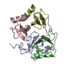
| ||||||||
|---|---|---|---|---|---|---|---|---|---|
| 1 |
| ||||||||
| Unit cell |
|
-Components
| #1: Protein | Mass: 7623.935 Da / Num. of mol.: 4 / Mutation: M26L Source method: isolated from a genetically manipulated source Source: (gene. exp.) Homo sapiens (human) / Plasmid: PBR-CRM-CTAP-MET20,LEU26 / Production host:  #2: Water | ChemComp-HOH / |  Mass: 18.015 Da / Num. of mol.: 265 / Source method: isolated from a natural source / Formula: H2O Mass: 18.015 Da / Num. of mol.: 265 / Source method: isolated from a natural source / Formula: H2OHas protein modification | Y | Sequence details | THE NUMBERING SCHEME FOR NAP-2 FOLLOWS HOMOLOGY ALIGNMENT WITH THE FIRST PAIR OF CYSTEINE RESIDUES ...THE NUMBERING SCHEME FOR NAP-2 FOLLOWS HOMOLOGY ALIGNMENT WITH THE FIRST PAIR OF CYSTEINE RESIDUES IN BOVINE PLATELET FACTOR FOUR. THE NUMBERING SCHEME IS SEQUENTIAL | |
|---|
-Experimental details
-Experiment
| Experiment | Method: X-RAY DIFFRACTION |
|---|
- Sample preparation
| Crystal | Density Matthews: 2.21 Å3/Da / Density % sol: 44.36 % | |||||||||||||||||||||||||
|---|---|---|---|---|---|---|---|---|---|---|---|---|---|---|---|---|---|---|---|---|---|---|---|---|---|---|
| Crystal grow | *PLUS Temperature: 22 ℃ / pH: 4.6 / Method: vapor diffusion, hanging drop | |||||||||||||||||||||||||
| Components of the solutions | *PLUS
|
-Data collection
| Radiation | Scattering type: x-ray |
|---|---|
| Radiation wavelength | Relative weight: 1 |
| Reflection | *PLUS Highest resolution: 1.9 Å / Lowest resolution: 7 Å / Num. obs: 17830 / % possible obs: 97 % / Num. measured all: 30511 / Rmerge(I) obs: 0.025 |
| Reflection shell | *PLUS Highest resolution: 1.9 Å / Lowest resolution: 2 Å / % possible obs: 47 % |
- Processing
| Software |
| ||||||||||||||||||||||||||||||||||||||||||||||||||||||||||||
|---|---|---|---|---|---|---|---|---|---|---|---|---|---|---|---|---|---|---|---|---|---|---|---|---|---|---|---|---|---|---|---|---|---|---|---|---|---|---|---|---|---|---|---|---|---|---|---|---|---|---|---|---|---|---|---|---|---|---|---|---|---|
| Refinement | Resolution: 1.9→7 Å Details: THE MUTATION M26L IS DESCRIBED AS M6L IN THE JRNL REFERENCE.
| ||||||||||||||||||||||||||||||||||||||||||||||||||||||||||||
| Refinement step | Cycle: LAST / Resolution: 1.9→7 Å
| ||||||||||||||||||||||||||||||||||||||||||||||||||||||||||||
| Refine LS restraints |
| ||||||||||||||||||||||||||||||||||||||||||||||||||||||||||||
| Refinement | *PLUS | ||||||||||||||||||||||||||||||||||||||||||||||||||||||||||||
| Solvent computation | *PLUS | ||||||||||||||||||||||||||||||||||||||||||||||||||||||||||||
| Displacement parameters | *PLUS Biso mean: 30.5 Å2 | ||||||||||||||||||||||||||||||||||||||||||||||||||||||||||||
| Refine LS restraints | *PLUS
|
