 Movie
Movie Controller

[English] 日本語
 Yorodumi
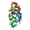
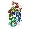
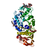
- PDB-1mwo: Crystal Structure Analysis of the Hyperthermostable Pyrocoocus wo... -
+ Open data

- Basic information
| Entry | Database: PDB / ID: 1mwo | ||||||
|---|---|---|---|---|---|---|---|
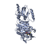
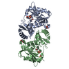
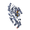
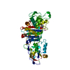
| Title | Crystal Structure Analysis of the Hyperthermostable Pyrocoocus woesei alpha-amylase | ||||||
 Components Components | alpha amylase | ||||||
 Keywords Keywords | HYDROLASE / (beta/alpha)8-barrel / alpha-amylase | ||||||
| Function / homology |  Function and homology information Function and homology informationalpha-amylase / alpha-amylase activity / carbohydrate metabolic process / calcium ion binding Similarity search - Function | ||||||
| Biological species |   Pyrococcus woesei (archaea) Pyrococcus woesei (archaea) | ||||||
| Method |  X-RAY DIFFRACTION / SYNCHROTRON / SIRAS / Resolution: 2.2 Å X-RAY DIFFRACTION / SYNCHROTRON / SIRAS / Resolution: 2.2 Å | ||||||
 Authors Authors | Linden, A. / Mayans, O. / Meyer-Klaucke, W. / Antranikian, G. / Wilmanns, M. | ||||||
 Citation Citation | Journal: J.Biol.Chem. / Year: 2003 Title: Differential Regulation of a Hyperthermophilic alpha-Amylase with a Novel (Ca,Zn) Two-metal Center by Zinc Authors: Linden, A. / Mayans, O. / Meyer-Klaucke, W. / Antranikian, G. / Wilmanns, M. | ||||||
| History |
|
- Structure visualization
| Structure viewer | Molecule: MolmilJmol/JSmol |
|---|
- Downloads & links
-Download
| PDBx/mmCIF format | 1mwo.cif.gz | 110.2 KB | Display | PDBx/mmCIF format |
|---|---|---|---|---|
| PDB format | pdb1mwo.ent.gz | 83.8 KB | Display | PDB format |
| PDBx/mmJSON format | 1mwo.json.gz | Tree view | PDBx/mmJSON format | |
| Others |  Other downloads Other downloads |
-Validation report
| Arichive directory | https://data.pdbj.org/pub/pdb/validation_reports/mw/1mwoftp://data.pdbj.org/pub/pdb/validation_reports/mw/1mwo | HTTPS FTP |
|---|
-Related structure data


-Links
 PDBj
PDBj

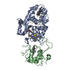
- Assembly
| Deposited unit | 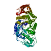
| ||||||||
|---|---|---|---|---|---|---|---|---|---|
| 1 |
| ||||||||
| Unit cell |
|
-Components
| #1: Protein | Mass: 50228.676 Da / Num. of mol.: 1 Source method: isolated from a genetically manipulated source Source: (gene. exp.) Pyrococcus woesei (archaea) / Plasmid: pET15b / Species (production host): Escherichia coli / Production host:  References: UniProt: O08452, UniProt: Q7LYT7*PLUS, alpha-amylase | ||||||
|---|---|---|---|---|---|---|---|
| #2: Chemical | ChemComp-ZN /   Mass: 65.409 Da / Num. of mol.: 6 / Source method: obtained synthetically / Formula: Zn Mass: 65.409 Da / Num. of mol.: 6 / Source method: obtained synthetically / Formula: Zn#3: Chemical | ChemComp-CA / |   Mass: 40.078 Da / Num. of mol.: 1 / Source method: obtained synthetically / Formula: Ca Mass: 40.078 Da / Num. of mol.: 1 / Source method: obtained synthetically / Formula: Ca#4: Water | ChemComp-HOH / |  Mass: 18.015 Da / Num. of mol.: 398 / Source method: isolated from a natural source / Formula: H2O Mass: 18.015 Da / Num. of mol.: 398 / Source method: isolated from a natural source / Formula: H2OHas protein modification | Y | |
-Experimental details
-Experiment
| Experiment | Method: X-RAY DIFFRACTION / Number of used crystals: 1 |
|---|
- Sample preparation
| Crystal | Density Matthews: 2.6 Å3/Da / Density % sol: 52.7 % | |||||||||||||||||||||||||||||||||||
|---|---|---|---|---|---|---|---|---|---|---|---|---|---|---|---|---|---|---|---|---|---|---|---|---|---|---|---|---|---|---|---|---|---|---|---|---|
| Crystal grow | Temperature: 292 K / Method: vapor diffusion, hanging drop / pH: 6.5 Details: PEG-MME 550, zinc sulfate, MES, pH 6.5, VAPOR DIFFUSION, HANGING DROP, temperature 292K | |||||||||||||||||||||||||||||||||||
| Crystal grow | *PLUS Temperature: 19 ℃ / Method: vapor diffusion, hanging drop | |||||||||||||||||||||||||||||||||||
| Components of the solutions | *PLUS
|
-Data collection
| Diffraction | Mean temperature: 100 K |
|---|---|
| Diffraction source | Source: SYNCHROTRON / Site: EMBL/DESY, HAMBURG  / Beamline: X11 / Wavelength: 0.91 Å / Beamline: X11 / Wavelength: 0.91 Å |
| Detector | Type: MARRESEARCH / Detector: CCD / Date: Jul 23, 2000 |
| Radiation | Monochromator: Triangular / Protocol: SINGLE WAVELENGTH / Monochromatic (M) / Laue (L): M / Scattering type: x-ray |
| Radiation wavelength | Wavelength: 0.91 Å / Relative weight: 1 |
| Reflection | Resolution: 2.2→20 Å / Num. all: 26004 / Num. obs: 26004 / % possible obs: 97.7 % / Observed criterion σ(F): 0 / Observed criterion σ(I): 0 / Redundancy: 3.4 % / Biso Wilson estimate: 27.1 Å2 / Rsym value: 0.06 / Net I/σ(I): 10.2 |
| Reflection shell | Highest resolution: 2.2 Å / Mean I/σ(I) obs: 3 / Num. unique all: 26004 / Rsym value: 0.178 / % possible all: 93.1 |
| Reflection | *PLUS Highest resolution: 2.2 Å / Rmerge(I) obs: 0.06 |
| Reflection shell | *PLUS Highest resolution: 2.22 Å / Lowest resolution: 2.28 Å / % possible obs: 93.1 % / Rmerge(I) obs: 0.178 |
- Processing
| Software |
| |||||||||||||||||||||||||
|---|---|---|---|---|---|---|---|---|---|---|---|---|---|---|---|---|---|---|---|---|---|---|---|---|---|---|
| Refinement | Method to determine structure: SIRAS / Resolution: 2.2→20 Å / Cross valid method: THROUGHOUT / σ(F): 0
| |||||||||||||||||||||||||
| Displacement parameters | Biso mean: 26.4 Å2
| |||||||||||||||||||||||||
| Refine analyze | Luzzati coordinate error obs: 0.263 Å | |||||||||||||||||||||||||
| Refinement step | Cycle: LAST / Resolution: 2.2→20 Å
| |||||||||||||||||||||||||
| Refine LS restraints |
| |||||||||||||||||||||||||
| Xplor file |
| |||||||||||||||||||||||||
| Refinement | *PLUS Rfactor Rwork: 0.19 | |||||||||||||||||||||||||
| Solvent computation | *PLUS | |||||||||||||||||||||||||
| Displacement parameters | *PLUS |
