 Movie
Movie Controller

[English] 日本語
 Yorodumi
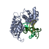
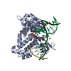
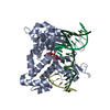
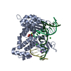
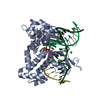
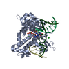
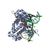
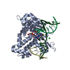
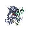
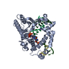
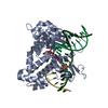
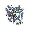
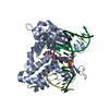
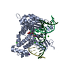
- PDB-1mq3: Human DNA Polymerase Beta Complexed With Gapped DNA Containing an... -
+ Open data

- Basic information
| Entry | Database: PDB / ID: 1mq3 | ||||||
|---|---|---|---|---|---|---|---|
| Title | Human DNA Polymerase Beta Complexed With Gapped DNA Containing an 8-oxo-7,8-dihydro-Guanine Template Paired with dCTP | ||||||
 Components Components |
| ||||||
 Keywords Keywords | TRANSFERASE/DNA / TRANSFERASE / DNA / TRANSFERASE-DNA COMPLEX | ||||||
| Function / homology |  Function and homology information Function and homology informationResolution of AP sites via the single-nucleotide replacement pathway / immunoglobulin heavy chain V-D-J recombination / Resolution of AP sites via the multiple-nucleotide patch replacement pathway / Abasic sugar-phosphate removal via the single-nucleotide replacement pathway / APEX1-Independent Resolution of AP Sites via the Single Nucleotide Replacement Pathway / Lyases; Carbon-oxygen lyases; Other carbon-oxygen lyases / pyrimidine dimer repair / POLB-Dependent Long Patch Base Excision Repair / homeostasis of number of cells / PCNA-Dependent Long Patch Base Excision Repair ...Resolution of AP sites via the single-nucleotide replacement pathway / immunoglobulin heavy chain V-D-J recombination / Resolution of AP sites via the multiple-nucleotide patch replacement pathway / Abasic sugar-phosphate removal via the single-nucleotide replacement pathway / APEX1-Independent Resolution of AP Sites via the Single Nucleotide Replacement Pathway / Lyases; Carbon-oxygen lyases; Other carbon-oxygen lyases / pyrimidine dimer repair / POLB-Dependent Long Patch Base Excision Repair / homeostasis of number of cells / PCNA-Dependent Long Patch Base Excision Repair / 5'-deoxyribose-5-phosphate lyase activity / response to hyperoxia / lymph node development / salivary gland morphogenesis / somatic hypermutation of immunoglobulin genes / spleen development / base-excision repair, gap-filling / DNA-(apurinic or apyrimidinic site) endonuclease activity / class I DNA-(apurinic or apyrimidinic site) endonuclease activity / DNA-(apurinic or apyrimidinic site) lyase / response to gamma radiation / spindle microtubule / base-excision repair / DNA-templated DNA replication / double-strand break repair via nonhomologous end joining / intrinsic apoptotic signaling pathway in response to DNA damage / neuron apoptotic process / DNA-directed DNA polymerase / microtubule binding / in utero embryonic development / damaged DNA binding / microtubule / DNA-directed DNA polymerase activity / response to ethanol / lyase activity / Ub-specific processing proteases / inflammatory response / DNA repair / DNA damage response / enzyme binding / protein-containing complex / nucleoplasm / metal ion binding / nucleus / cytoplasm Similarity search - Function | ||||||
| Biological species |  Homo sapiens (human) Homo sapiens (human) | ||||||
| Method |  X-RAY DIFFRACTION / MOLECULAR REPLACEMENT / Resolution: 2.8 Å X-RAY DIFFRACTION / MOLECULAR REPLACEMENT / Resolution: 2.8 Å | ||||||
 Authors Authors | Krahn, J.M. / Beard, W.A. / Miller, H. / Grollman, A.P. / Wilson, S.H. | ||||||
 Citation Citation | Journal: Structure / Year: 2003 Title: Structure of DNA Polymerase beta with the Mutagenic DNA Lesion 8-oxodeoxyguanine Reveals Structural Insights into its Coding Potential Authors: Krahn, J.M. / Beard, W.A. / Miller, H. / Grollman, A.P. / Wilson, S.H. | ||||||
| History |
|
- Structure visualization
| Structure viewer | Molecule: MolmilJmol/JSmol |
|---|
- Downloads & links
-Download
| PDBx/mmCIF format | 1mq3.cif.gz | 101.7 KB | Display | PDBx/mmCIF format |
|---|---|---|---|---|
| PDB format | pdb1mq3.ent.gz | 72.8 KB | Display | PDB format |
| PDBx/mmJSON format | 1mq3.json.gz | Tree view | PDBx/mmJSON format | |
| Others |  Other downloads Other downloads |
-Validation report
| Arichive directory | https://data.pdbj.org/pub/pdb/validation_reports/mq/1mq3ftp://data.pdbj.org/pub/pdb/validation_reports/mq/1mq3 | HTTPS FTP |
|---|
-Related structure data
| Related structure data | 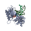 1mq2C 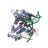 1bpyS S: Starting model for refinement C: citing same article ( |
|---|---|
| Similar structure data |


-Links
 PDBj
PDBj


- Assembly
| Deposited unit | 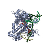
| ||||||||
|---|---|---|---|---|---|---|---|---|---|
| 1 |
| ||||||||
| Unit cell |
|
-Components
-DNA chain , 3 types, 3 molecules TPD
| #1: DNA chain | Mass: 4885.157 Da / Num. of mol.: 1 / Source method: obtained synthetically |
|---|---|
| #2: DNA chain | Mass: 3045.005 Da / Num. of mol.: 1 / Source method: obtained synthetically |
| #3: DNA chain | Mass: 1536.035 Da / Num. of mol.: 1 / Source method: obtained synthetically |
-Protein , 1 types, 1 molecules A
| #4: Protein | Mass: 38241.672 Da / Num. of mol.: 1 Source method: isolated from a genetically manipulated source Source: (gene. exp.) Homo sapiens (human) / Production host:  |
|---|
-Non-polymers , 4 types, 82 molecules 


| #5: Chemical | ChemComp-MG /  Mass: 24.305 Da / Num. of mol.: 1 / Source method: obtained synthetically / Formula: Mg Mass: 24.305 Da / Num. of mol.: 1 / Source method: obtained synthetically / Formula: Mg | ||||
|---|---|---|---|---|---|
| #6: Chemical |  Mass: 22.990 Da / Num. of mol.: 2 / Source method: obtained synthetically / Formula: Na Mass: 22.990 Da / Num. of mol.: 2 / Source method: obtained synthetically / Formula: Na#7: Chemical | ChemComp-DCP / |  Mass: 467.157 Da / Num. of mol.: 1 / Source method: obtained synthetically / Formula: C9H16N3O13P3 Mass: 467.157 Da / Num. of mol.: 1 / Source method: obtained synthetically / Formula: C9H16N3O13P3#8: Water | ChemComp-HOH / | Mass: 18.015 Da / Num. of mol.: 78 / Source method: isolated from a natural source / Formula: H2O |
-Experimental details
-Experiment
| Experiment | Method: X-RAY DIFFRACTION / Number of used crystals: 1 |
|---|
- Sample preparation
| Crystal | Density Matthews: 2.22 Å3/Da / Density % sol: 44.55 % | ||||||||||||||||||||||||||||||||||||
|---|---|---|---|---|---|---|---|---|---|---|---|---|---|---|---|---|---|---|---|---|---|---|---|---|---|---|---|---|---|---|---|---|---|---|---|---|---|
| Crystal grow | Temperature: 291 K / pH: 7.5 Details: 14% PEG 3350, 350 mM sodium acetate and 50 mM imidazole at 18 degrees (C), pH 7.5, temperature 291.0K | ||||||||||||||||||||||||||||||||||||
| Components of the solutions |
| ||||||||||||||||||||||||||||||||||||
| Crystal grow | *PLUS Temperature: 18 ℃ / pH: 7 / Method: vapor diffusion, sitting drop | ||||||||||||||||||||||||||||||||||||
| Components of the solutions | *PLUS
|
-Data collection
| Diffraction | Mean temperature: 200 K |
|---|---|
| Diffraction source | Source: ROTATING ANODE / Type: RIGAKU RU300 / Wavelength: 1.5418 |
| Detector | Type: RIGAKU RAXIS IV / Detector: IMAGE PLATE |
| Radiation | Protocol: SINGLE WAVELENGTH / Monochromatic (M) / Laue (L): M / Scattering type: x-ray |
| Radiation wavelength | Wavelength: 1.5418 Å / Relative weight: 1 |
| Reflection | Resolution: 2.8→20 Å / Num. obs: 10209 / % possible obs: 97.8 % / Observed criterion σ(I): -3 / Redundancy: 2.51 % / Rmerge(I) obs: 0.108 / Net I/σ(I): 5.5 |
| Reflection shell | Resolution: 2.8→2.93 Å / Rmerge(I) obs: 0.339 / Mean I/σ(I) obs: 1.9 / % possible all: 95.9 |
| Reflection | *PLUS Num. all: 25622 |
| Reflection shell | *PLUS % possible obs: 95.9 % / Num. possible: 1769 / Num. unique obs: 981 |
- Processing
| Software |
| |||||||||||||||||||||||||
|---|---|---|---|---|---|---|---|---|---|---|---|---|---|---|---|---|---|---|---|---|---|---|---|---|---|---|
| Refinement | Method to determine structure: MOLECULAR REPLACEMENT Starting model: 1BPY Resolution: 2.8→20 Å / Cross valid method: THROUGHOUT / σ(F): 0 / Stereochemistry target values: Engh & Huber Details: REFINEMENT PARAMETERS FOR 8-OXO-G ARE BASED ON SEVERAL SMALL MOLECULE OBSERVATIONS. DIHEDRAL RESTRAINTS FROM DNA-RNA.PARAM WERE DISABLED TO ALLOW FOR DEVIATIONS FROM CANONICAL B-FORM VALUES, ...Details: REFINEMENT PARAMETERS FOR 8-OXO-G ARE BASED ON SEVERAL SMALL MOLECULE OBSERVATIONS. DIHEDRAL RESTRAINTS FROM DNA-RNA.PARAM WERE DISABLED TO ALLOW FOR DEVIATIONS FROM CANONICAL B-FORM VALUES, WHICH HAVE BEEN OBSERVED IN HIGH-RESOLUTION DATA FROM SIMILAR POLYMERASE BETA DNA COMPLEXES.
| |||||||||||||||||||||||||
| Solvent computation | Bsol: 10 Å2 / ksol: 0.2998 e/Å3 | |||||||||||||||||||||||||
| Displacement parameters | Biso mean: 22.42 Å2
| |||||||||||||||||||||||||
| Refinement step | Cycle: LAST / Resolution: 2.8→20 Å
| |||||||||||||||||||||||||
| Refine LS restraints |
| |||||||||||||||||||||||||
| Xplor file |
| |||||||||||||||||||||||||
| Refinement | *PLUS % reflection Rfree: 5 % | |||||||||||||||||||||||||
| Solvent computation | *PLUS | |||||||||||||||||||||||||
| Displacement parameters | *PLUS | |||||||||||||||||||||||||
| Refine LS restraints | *PLUS Type: c_angle_deg / Dev ideal: 1.1 |
