 ムービー
ムービー コントローラー


+ データを開く

- 基本情報
基本情報
| 登録情報 | データベース: PDB / ID: 1mpe | ||||||
|---|---|---|---|---|---|---|---|
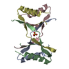
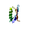
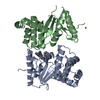
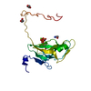
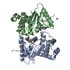
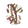
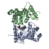
| タイトル | Ensemble of 20 structures of the tetrameric mutant of the B1 domain of streptococcal protein G | ||||||
 要素 要素 | Immunoglobulin G binding protein G | ||||||
 キーワード キーワード | PROTEIN BINDING / strand-exchanged tetramer / channel | ||||||
| 機能・相同性 |  機能・相同性情報 機能・相同性情報 | ||||||
| 生物種 |  Streptococcus sp. 'group G' (バクテリア) Streptococcus sp. 'group G' (バクテリア) | ||||||
| 手法 | 溶液NMR / simulated annealing | ||||||
| Model type details | minimized average | ||||||
 データ登録者 データ登録者 | Frank, M.K. / Dyda, F. / Dobrodumov, A. / Gronenborn, A.M. | ||||||
 引用 引用 | ジャーナル: Nat.Struct.Biol. / 年: 2002 タイトル: Core mutations switch monomeric protein GB1 into an intertwined tetramer. 著者: Kirsten Frank, M. / Dyda, F. / Dobrodumov, A. / Gronenborn, A.M. #1: ジャーナル: Science / 年: 1991タイトル: A Novel, Highly Stable Fold of the Immunoglobulin Binding Domain of Streptococcal Protein G. 著者: Gronenborn, A.M. / Filpula, D.R. / Essig, N.Z. / Achari, A. / Whitlow, M. / Wingfield, P.T. / Clore, G.M. #2: ジャーナル: FEBS Lett. / 年: 1996タイトル: Core Mutants of the Immunoglobulin Binding Domain of Streptococcal Protein G: Stability and Structural Integrity 著者: Gronenborn, A.M. / Frank, M.K. / Clore, G.M. | ||||||
| 履歴 |
|
- 構造の表示
構造の表示
| 構造ビューア | 分子: MolmilJmol/JSmol |
|---|
- ダウンロードとリンク
-ダウンロード
| PDBx/mmCIF形式 | 1mpe.cif.gz | 1.4 MB | 表示 | PDBx/mmCIF形式 |
|---|---|---|---|---|
| PDB形式 | pdb1mpe.ent.gz | 1.2 MB | 表示 | PDB形式 |
| PDBx/mmJSON形式 | 1mpe.json.gz | ツリー表示 | PDBx/mmJSON形式 | |
| その他 |  その他のダウンロード その他のダウンロード |
-検証レポート
| アーカイブディレクトリ | https://data.pdbj.org/pub/pdb/validation_reports/mp/1mpeftp://data.pdbj.org/pub/pdb/validation_reports/mp/1mpe | HTTPS FTP |
|---|
-関連構造データ


-リンク
 PDBj
PDBj

- 集合体
集合体
| 登録構造単位 | 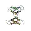
| |||||||||
|---|---|---|---|---|---|---|---|---|---|---|
| 1 |
| |||||||||
| NMR アンサンブル |
|
-要素
| #1: 抗体 | 分子量: 6302.931 Da / 分子数: 4 / 断片: B1 domain, sequence database residues 228-282 / 変異: T2Q, L5V, A26F, F30V, Y33F, A34F / 由来タイプ: 組換発現 由来: (組換発現) Streptococcus sp. 'group G' (バクテリア)遺伝子: SPG / プラスミド: pet11a / 発現宿主: |
|---|
-実験情報
-実験
| 実験 | 手法: 溶液NMR | ||||||||||||||||||||||||||||
|---|---|---|---|---|---|---|---|---|---|---|---|---|---|---|---|---|---|---|---|---|---|---|---|---|---|---|---|---|---|
| NMR実験 |
| ||||||||||||||||||||||||||||
| NMR実験の詳細 | Text: T1,T2, and heteronuclear NOEs collected. Dipolar couplings (N-H) were collected as well. Dipolar coupling and chemical shifts from residues with heteronuclear NOE below 0.6 were not used. |
- 試料調製
試料調製
| 詳細 |
| |||||||||||||||
|---|---|---|---|---|---|---|---|---|---|---|---|---|---|---|---|---|
| 試料状態 | イオン強度: 50 mM sodium phosphate, 0.02% sodium azide pH: 5.45 / 圧: 1 atm / 温度: 313 K | |||||||||||||||
| 結晶化 | *PLUS 手法: other / 詳細: NMR |
-NMR測定
| 放射 | プロトコル: SINGLE WAVELENGTH / 単色(M)・ラウエ(L): M | |||||||||||||||||||||||||
|---|---|---|---|---|---|---|---|---|---|---|---|---|---|---|---|---|---|---|---|---|---|---|---|---|---|---|
| 放射波長 | 相対比: 1 | |||||||||||||||||||||||||
| NMRスペクトロメーター |
|
- 解析
解析
| NMR software |
| ||||||||||||||||||||||||
|---|---|---|---|---|---|---|---|---|---|---|---|---|---|---|---|---|---|---|---|---|---|---|---|---|---|
| 精密化 | 手法: simulated annealing / ソフトェア番号: 1 詳細: NMR constraints (per monomer) 822 NOE-derived distance restraints (534 intramonomer, 288 intermonomer) 42 distance restraints from hydrogen bonds (20 intramonomer, 22 intermonomer) 30 J- ...詳細: NMR constraints (per monomer) 822 NOE-derived distance restraints (534 intramonomer, 288 intermonomer) 42 distance restraints from hydrogen bonds (20 intramonomer, 22 intermonomer) 30 J-coupling restraints, 33 Residual dipolar couplings (HN) 66 backbone dihedral angle restraints 31 sidechain dihedral angle restraints | ||||||||||||||||||||||||
| 代表構造 | 選択基準: minimized average structure | ||||||||||||||||||||||||
| NMRアンサンブル | コンフォーマー選択の基準: structures with least restraint violations and phi, psi angles of residue 46 in allowed region of Ramachrandran plot 計算したコンフォーマーの数: 100 / 登録したコンフォーマーの数: 21 |
 NMRPipe