 Movie
Movie Controller


+ Open data

- Basic information
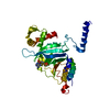
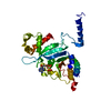
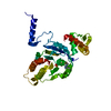
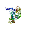
| Entry | Database: PDB / ID: 1mo5 | ||||||
|---|---|---|---|---|---|---|---|
| Title | RECA-ATP-GAMMA-S-MG COMPLEX | ||||||
 Components Components | RecA | ||||||
 Keywords Keywords | HYDROLASE / RECOMBINATION / DNA-REPAIR / Structural Genomics / PSI / Protein Structure Initiative / TB Structural Genomics Consortium / TBSGC | ||||||
| Function / homology |  Function and homology information Function and homology informationDNA strand invasion / intron homing / intein-mediated protein splicing / UV protection / DNA strand exchange activity / SOS response / recombinational repair / ATP-dependent DNA damage sensor activity / ATP-dependent activity, acting on DNA / DNA endonuclease activity ...DNA strand invasion / intron homing / intein-mediated protein splicing / UV protection / DNA strand exchange activity / SOS response / recombinational repair / ATP-dependent DNA damage sensor activity / ATP-dependent activity, acting on DNA / DNA endonuclease activity / manganese ion binding / single-stranded DNA binding / endonuclease activity / damaged DNA binding / Hydrolases; Acting on ester bonds / response to antibiotic / DNA repair / DNA damage response / magnesium ion binding / ATP hydrolysis activity / ATP binding / cytosol / cytoplasm Similarity search - Function | ||||||
| Biological species |   Mycobacterium tuberculosis (bacteria) Mycobacterium tuberculosis (bacteria) | ||||||
| Method |  X-RAY DIFFRACTION / MOLECULAR REPLACEMENT / Resolution: 3.25 Å X-RAY DIFFRACTION / MOLECULAR REPLACEMENT / Resolution: 3.25 Å | ||||||
 Authors Authors | Datta, S. / Ganesh, N. / Chandra, N.R. / TB Structural Genomics Consortium (TBSGC) | ||||||
 Citation Citation | Journal: Proteins / Year: 2003 Title: Structural studies on MtRecA-nucleotide complexes: Insights into DNA and nucleotide binding and the structural signature of NTP recognition Authors: Datta, S. / Ganesh, N. / Chandra, N.R. / Muniyappa, K. / Vijayan, M. #1: Journal: Nucleic Acids Res. / Year: 2000Title: Crystal Structures of Mycobacterium Tuberculosis Reca and its Complex with Adp-Alf4: Implications For Decreased ATPase Activity and Molecular Aggregation Authors: Datta, S. / Prabu, M. / Vaze, M.B. / Ganesh, N. / Chandra, N.R. / Muniyappa, K. / Vijayan, M. #2: Journal: Biochemistry / Year: 1996 Title: Functional Characterization of the Precursor and Spliced Forms of Reca Protein of Mycobacterium Tuberculosis. Authors: Ajay Kumar, R. / Vaze, M. / Chandra, N.R. / Vijayan, M. / Muniyappa, K. | ||||||
| History |
|
- Structure visualization
| Structure viewer | Molecule: MolmilJmol/JSmol |
|---|
- Downloads & links
-Download
| PDBx/mmCIF format | 1mo5.cif.gz | 70.7 KB | Display | PDBx/mmCIF format |
|---|---|---|---|---|
| PDB format | pdb1mo5.ent.gz | 51.7 KB | Display | PDB format |
| PDBx/mmJSON format | 1mo5.json.gz | Tree view | PDBx/mmJSON format | |
| Others |  Other downloads Other downloads |
-Validation report
| Arichive directory | https://data.pdbj.org/pub/pdb/validation_reports/mo/1mo5ftp://data.pdbj.org/pub/pdb/validation_reports/mo/1mo5 | HTTPS FTP |
|---|
-Related structure data
| Related structure data |  1mo3C  1mo4C  1mo6C 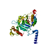 1g19S C: citing same article ( S: Starting model for refinement |
|---|---|
| Similar structure data | |
| Other databases |


-Links
 PDBj
PDBj


- Assembly
| Deposited unit | 
| ||||||||
|---|---|---|---|---|---|---|---|---|---|
| 1 |
| ||||||||
| Unit cell |
|
-Components
| #1: Protein | Mass: 37222.281 Da / Num. of mol.: 1 Source method: isolated from a genetically manipulated source Source: (gene. exp.) Mycobacterium tuberculosis (bacteria) / Plasmid: pEJ135 / Production host: References: UniProt: P0A5U4, UniProt: P9WHJ3*PLUS, EC: 3.4.99.37 |
|---|---|
| #2: Chemical | ChemComp-AGS /   Mass: 523.247 Da / Num. of mol.: 1 / Source method: obtained synthetically / Formula: C10H16N5O12P3S / Comment: ATP-gamma-S, energy-carrying molecule analogue*YM Mass: 523.247 Da / Num. of mol.: 1 / Source method: obtained synthetically / Formula: C10H16N5O12P3S / Comment: ATP-gamma-S, energy-carrying molecule analogue*YM |
| #3: Water | ChemComp-HOH /  Mass: 18.015 Da / Num. of mol.: 61 / Source method: isolated from a natural source / Formula: H2O Mass: 18.015 Da / Num. of mol.: 61 / Source method: isolated from a natural source / Formula: H2O |
| Sequence details | THIS STRUCTURE CORRESPONDS TO THE MATURE MTRECA, A 38 KD POLYPEPTIDE CHAIN WHICH IS AN AUTOSPLICED ...THIS STRUCTURE CORRESPOND |
-Experimental details
-Experiment
| Experiment | Method: X-RAY DIFFRACTION / Number of used crystals: 1 |
|---|
- Sample preparation
| Crystal | Density Matthews: 3.213 Å3/Da / Density % sol: 60.99 % | ||||||||||||||||||||||||||||||||||||||||||
|---|---|---|---|---|---|---|---|---|---|---|---|---|---|---|---|---|---|---|---|---|---|---|---|---|---|---|---|---|---|---|---|---|---|---|---|---|---|---|---|---|---|---|---|
| Crystal grow | Temperature: 293 K / Method: vapor diffusion, hanging drop / pH: 7 Details: PEG 4000, TRIS-ACETATE, NACL, pH 7.0, VAPOR DIFFUSION, HANGING DROP, temperature 293K | ||||||||||||||||||||||||||||||||||||||||||
| Crystal grow | *PLUS pH: 7 | ||||||||||||||||||||||||||||||||||||||||||
| Components of the solutions | *PLUS
|
-Data collection
| Diffraction | Mean temperature: 293 K |
|---|---|
| Diffraction source | Source: ROTATING ANODE / Type: RIGAKU RU200 / Wavelength: 1.5418 / Wavelength: 1.5418 Å |
| Detector | Type: MARRESEARCH / Detector: IMAGE PLATE / Date: Oct 22, 2001 |
| Radiation | Protocol: SINGLE WAVELENGTH / Monochromatic (M) / Laue (L): M / Scattering type: x-ray |
| Radiation wavelength | Wavelength: 1.5418 Å / Relative weight: 1 |
| Reflection | Resolution: 3.25→30 Å / Num. all: 7435 / Num. obs: 7435 / % possible obs: 96.6 % / Observed criterion σ(F): 0 / Observed criterion σ(I): 0 / Redundancy: 3.7 % / Rmerge(I) obs: 0.093 / Net I/σ(I): 7.7 |
| Reflection shell | Resolution: 3.25→3.37 Å / Redundancy: 3.5 % / Rmerge(I) obs: 0.314 / Mean I/σ(I) obs: 2.9 / Num. unique all: 720 / % possible all: 95.9 |
| Reflection | *PLUS Num. measured all: 30714 |
| Reflection shell | *PLUS % possible obs: 95.9 % |
- Processing
| Software |
| ||||||||||||||||||||||||||||||||||||||||||||||||||||||||||||
|---|---|---|---|---|---|---|---|---|---|---|---|---|---|---|---|---|---|---|---|---|---|---|---|---|---|---|---|---|---|---|---|---|---|---|---|---|---|---|---|---|---|---|---|---|---|---|---|---|---|---|---|---|---|---|---|---|---|---|---|---|---|
| Refinement | Method to determine structure: MOLECULAR REPLACEMENT Starting model: PDB ENTRY 1G19 Resolution: 3.25→30 Å / Rfactor Rfree error: 0.01 / Isotropic thermal model: GROUP / Cross valid method: THROUGHOUT / σ(F): 0 / Stereochemistry target values: Engh & Huber
| ||||||||||||||||||||||||||||||||||||||||||||||||||||||||||||
| Solvent computation | Solvent model: FLAT MODEL / Bsol: 88.8817 Å2 / ksol: 0.346201 e/Å3 | ||||||||||||||||||||||||||||||||||||||||||||||||||||||||||||
| Displacement parameters | Biso mean: 40.2 Å2
| ||||||||||||||||||||||||||||||||||||||||||||||||||||||||||||
| Refine analyze |
| ||||||||||||||||||||||||||||||||||||||||||||||||||||||||||||
| Refinement step | Cycle: LAST / Resolution: 3.25→30 Å
| ||||||||||||||||||||||||||||||||||||||||||||||||||||||||||||
| Refine LS restraints |
| ||||||||||||||||||||||||||||||||||||||||||||||||||||||||||||
| LS refinement shell | Resolution: 3.25→3.45 Å / Rfactor Rfree error: 0.032 / Total num. of bins used: 6
| ||||||||||||||||||||||||||||||||||||||||||||||||||||||||||||
| Xplor file |
| ||||||||||||||||||||||||||||||||||||||||||||||||||||||||||||
| Refinement | *PLUS Rfactor Rwork: 0.187 | ||||||||||||||||||||||||||||||||||||||||||||||||||||||||||||
| Solvent computation | *PLUS | ||||||||||||||||||||||||||||||||||||||||||||||||||||||||||||
| Displacement parameters | *PLUS | ||||||||||||||||||||||||||||||||||||||||||||||||||||||||||||
| Refine LS restraints | *PLUS
|
