 Movie
Movie Controller


+ Open data

- Basic information
| Entry | Database: PDB / ID: 1mn2 | |||||||||
|---|---|---|---|---|---|---|---|---|---|---|
| Title | MANGANESE PEROXIDASE SUBSTRATE BINDING SITE MUTANT E35Q, D179N | |||||||||
 Components Components | MANGANESE PEROXIDASE | |||||||||
 Keywords Keywords | PEROXIDASE / DONOR: H2O2 OXIDOREDUCTASE / HEME ENZYME | |||||||||
| Function / homology |  Function and homology information Function and homology informationmanganese peroxidase / manganese peroxidase activity / lignin catabolic process / response to reactive oxygen species / hydrogen peroxide catabolic process / cellular response to oxidative stress / heme binding / extracellular region / metal ion binding Similarity search - Function | |||||||||
| Biological species |  Phanerochaete chrysosporium (fungus) Phanerochaete chrysosporium (fungus) | |||||||||
| Method |  X-RAY DIFFRACTION / DIFFERENCE FOURIER / Resolution: 2 Å X-RAY DIFFRACTION / DIFFERENCE FOURIER / Resolution: 2 Å | |||||||||
 Authors Authors | Sundaramoorthy, M. / Poulos, T.L. | |||||||||
 Citation Citation | Journal: J.Biol.Chem. / Year: 1997 Title: Crystal structures of substrate binding site mutants of manganese peroxidase. Authors: Sundaramoorthy, M. / Kishi, K. / Gold, M.H. / Poulos, T.L. #1: Journal: J.Biol.Chem. / Year: 1994Title: The Crystal Structure of Manganese Peroxidase from Phanerochaete Chrysosporium at 2.06-A Resolution Authors: Sundaramoorthy, M. / Kishi, K. / Gold, M.H. / Poulos, T.L. | |||||||||
| History |
|
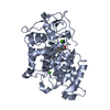
- Structure visualization
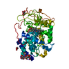
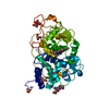
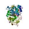
| Structure viewer | Molecule: MolmilJmol/JSmol |
|---|
- Downloads & links
-Download
| PDBx/mmCIF format | 1mn2.cif.gz | 86.6 KB | Display | PDBx/mmCIF format |
|---|---|---|---|---|
| PDB format | pdb1mn2.ent.gz | 64.3 KB | Display | PDB format |
| PDBx/mmJSON format | 1mn2.json.gz | Tree view | PDBx/mmJSON format | |
| Others |  Other downloads Other downloads |
-Validation report
| Arichive directory | https://data.pdbj.org/pub/pdb/validation_reports/mn/1mn2ftp://data.pdbj.org/pub/pdb/validation_reports/mn/1mn2 | HTTPS FTP |
|---|
-Related structure data


-Links
 PDBj
PDBj
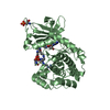
- Assembly
| Deposited unit | 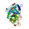
| ||||||||
|---|---|---|---|---|---|---|---|---|---|
| 1 |
| ||||||||
| Unit cell |
|
-Components
| #1: Protein | Mass: 37481.000 Da / Num. of mol.: 1 / Mutation: E35Q, D179N Source method: isolated from a genetically manipulated source Source: (gene. exp.) Phanerochaete chrysosporium (fungus) / Strain: OGC101 / Description: WOOD-ROTTING FUNGUS / Cellular location: EXTRACELLULAR / Gene: MNP1 / Variant: OGC107-1 / Plasmid: PUC18 / Gene (production host): MNP1 / Production host: Phanerochaete chrysosporium (fungus) / Strain (production host): OGC107-1 (ADE1) / Variant (production host): D179N-6 / References: UniProt: Q02567, manganese peroxidase | ||||||
|---|---|---|---|---|---|---|---|
| #2: Polysaccharide | 2-acetamido-2-deoxy-beta-D-glucopyranose-(1-4)-2-acetamido-2-deoxy-beta-D-glucopyranose Source method: isolated from a genetically manipulated source | ||||||
| #3: Chemical |   Mass: 40.078 Da / Num. of mol.: 2 / Source method: obtained synthetically / Formula: Ca Mass: 40.078 Da / Num. of mol.: 2 / Source method: obtained synthetically / Formula: Ca#4: Chemical | ChemComp-HEM / |   Mass: 616.487 Da / Num. of mol.: 1 / Source method: obtained synthetically / Formula: C34H32FeN4O4 Mass: 616.487 Da / Num. of mol.: 1 / Source method: obtained synthetically / Formula: C34H32FeN4O4#5: Water | ChemComp-HOH / |  Mass: 18.015 Da / Num. of mol.: 256 / Source method: isolated from a natural source / Formula: H2O Mass: 18.015 Da / Num. of mol.: 256 / Source method: isolated from a natural source / Formula: H2OHas protein modification | Y | |
-Experimental details
-Experiment
| Experiment | Method: X-RAY DIFFRACTION / Number of used crystals: 1 |
|---|
- Sample preparation
| Crystal | Density Matthews: 2.66 Å3/Da / Density % sol: 53.73 % Description: NATIVE STRUCTURE (CODE :1MNP) WAS USED AS THE STARTING MODEL | ||||||||||||||||||||||||||||||
|---|---|---|---|---|---|---|---|---|---|---|---|---|---|---|---|---|---|---|---|---|---|---|---|---|---|---|---|---|---|---|---|
| Crystal grow | pH: 6.5 Details: 30% PEG 8000, 0.2 M AMMONIUM SULFATE, 0.1 M SODIUM CACODYLATE BUFFER, PH 6.5 | ||||||||||||||||||||||||||||||
| Crystal grow | *PLUS Method: vapor diffusion, hanging drop | ||||||||||||||||||||||||||||||
| Components of the solutions | *PLUS
|
-Data collection
| Diffraction | Mean temperature: 295 K |
|---|---|
| Diffraction source | Source: ROTATING ANODE / Type: SIEMENS / Wavelength: 1.5418 |
| Detector | Type: SIEMENS / Detector: AREA DETECTOR / Date: Oct 3, 1995 / Details: SUPPER MIRRORS |
| Radiation | Monochromatic (M) / Laue (L): M / Scattering type: x-ray |
| Radiation wavelength | Wavelength: 1.5418 Å / Relative weight: 1 |
| Reflection | Highest resolution: 2 Å / Num. obs: 27424 / % possible obs: 98 % / Observed criterion σ(I): 1 / Redundancy: 2.9 % / Rsym value: 0.106 / Net I/σ(I): 13.6 |
| Reflection shell | Resolution: 2→2.2 Å / Redundancy: 2.1 % / Mean I/σ(I) obs: 1.9 / Rsym value: 0.294 / % possible all: 98 |
| Reflection | *PLUS Num. measured all: 91040 / Rmerge(I) obs: 0.1061 |
- Processing
| Software |
| ||||||||||||||||||||||||||||||||||||||||||||||||||||||||||||
|---|---|---|---|---|---|---|---|---|---|---|---|---|---|---|---|---|---|---|---|---|---|---|---|---|---|---|---|---|---|---|---|---|---|---|---|---|---|---|---|---|---|---|---|---|---|---|---|---|---|---|---|---|---|---|---|---|---|---|---|---|---|
| Refinement | Method to determine structure: DIFFERENCE FOURIER / Resolution: 2→8 Å / σ(F): 2 /
| ||||||||||||||||||||||||||||||||||||||||||||||||||||||||||||
| Refinement step | Cycle: LAST / Resolution: 2→8 Å
| ||||||||||||||||||||||||||||||||||||||||||||||||||||||||||||
| Refine LS restraints |
| ||||||||||||||||||||||||||||||||||||||||||||||||||||||||||||
| Xplor file |
| ||||||||||||||||||||||||||||||||||||||||||||||||||||||||||||
| Software | *PLUS Name: X-PLOR / Version: 3.8 / Classification: refinement | ||||||||||||||||||||||||||||||||||||||||||||||||||||||||||||
| Refinement | *PLUS Num. reflection all: 25117 | ||||||||||||||||||||||||||||||||||||||||||||||||||||||||||||
| Solvent computation | *PLUS | ||||||||||||||||||||||||||||||||||||||||||||||||||||||||||||
| Displacement parameters | *PLUS |
