 Movie
Movie Controller


+ Open data

- Basic information
| Entry | Database: PDB / ID: 1mab | ||||||
|---|---|---|---|---|---|---|---|
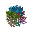
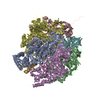
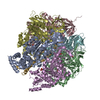
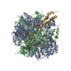
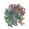
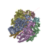
| Title | RAT LIVER F1-ATPASE | ||||||
 Components Components | (PROTEIN (F1-ATPASE ...) x 3 | ||||||
 Keywords Keywords | HYDROLASE / ATP SYNTHASE / FOF1-ATPASE / OXIDATIVE PHOSPHORYLATION / MITOCHONDRIA | ||||||
| Function / homology |  Function and homology information Function and homology informationlipoprotein particle receptor activity / Mitochondrial protein import / Formation of ATP by chemiosmotic coupling / Cristae formation / Mitochondrial protein degradation / negative regulation of cell adhesion involved in substrate-bound cell migration / response to 3,3',5-triiodo-L-thyronine / cold acclimation / response to curcumin / angiostatin binding ...lipoprotein particle receptor activity / Mitochondrial protein import / Formation of ATP by chemiosmotic coupling / Cristae formation / Mitochondrial protein degradation / negative regulation of cell adhesion involved in substrate-bound cell migration / response to 3,3',5-triiodo-L-thyronine / cold acclimation / response to curcumin / angiostatin binding / cellular response to peptide / ATP biosynthetic process / response to manganese ion / cellular response to interleukin-7 / response to muscle activity / negative regulation of endothelial cell proliferation / proton motive force-driven ATP synthesis / proton motive force-driven mitochondrial ATP synthesis / mitochondrial nucleoid / proton-transporting ATPase activity, rotational mechanism / MHC class I protein binding / positive regulation of blood vessel endothelial cell migration / cellular response to dexamethasone stimulus / H+-transporting two-sector ATPase / proton-transporting ATP synthase complex / proton-transporting ATP synthase activity, rotational mechanism / cellular response to nitric oxide / receptor-mediated endocytosis / proton transmembrane transport / regulation of intracellular pH / lipid metabolic process / liver development / ADP binding / mitochondrial membrane / protease binding / angiogenesis / response to ethanol / mitochondrial inner membrane / membrane raft / calcium ion binding / cell surface / ATP hydrolysis activity / mitochondrion / ATP binding / membrane / metal ion binding / plasma membrane Similarity search - Function | ||||||
| Biological species |  | ||||||
| Method |  X-RAY DIFFRACTION / MIR / Resolution: 2.8 Å X-RAY DIFFRACTION / MIR / Resolution: 2.8 Å | ||||||
 Authors Authors | Bianchet, M.A. / Amzel, L.M. | ||||||
 Citation Citation | Journal: Proc.Natl.Acad.Sci.USA / Year: 1998 Title: The 2.8-A structure of rat liver F1-ATPase: configuration of a critical intermediate in ATP synthesis/hydrolysis. Authors: Bianchet, M.A. / Hullihen, J. / Pedersen, P.L. / Amzel, L.M. #1: Journal: Biochim.Biophys.Acta / Year: 1994Title: The Three-Dimensional Structure of Rat Liver Mitochondria F1-ATPase: X-Ray Diffraction Studies Authors: Bianchet, M.A. / Medjahed, D. / Hullihen, J. / Pedersen, P.L. / Amzel, L.M. #2: Journal: J.Bioenerg.Biomembr. / Year: 1992Title: Quaternary Structure of ATP Synthases: Symmetry and Asymmetry in the F1 Moiety Authors: Amzel, L.M. / Bianchet, M.A. / Pedersen, P.L. #3: Journal: J.Biol.Chem. / Year: 1991Title: Quaternary Structure of ATP Synthases: Symmetry and Asymmetry in the F1 Moiety Authors: Bianchet, M.A. / Ysernhullihen, J. / Pedersen, P.L. / Amzel, L.M. | ||||||
| History |
|
- Structure visualization
| Structure viewer | Molecule: MolmilJmol/JSmol |
|---|
- Downloads & links
-Download
| PDBx/mmCIF format | 1mab.cif.gz | 226.3 KB | Display | PDBx/mmCIF format |
|---|---|---|---|---|
| PDB format | pdb1mab.ent.gz | 176.4 KB | Display | PDB format |
| PDBx/mmJSON format | 1mab.json.gz | Tree view | PDBx/mmJSON format | |
| Others |  Other downloads Other downloads |
-Validation report
| Arichive directory | https://data.pdbj.org/pub/pdb/validation_reports/ma/1mabftp://data.pdbj.org/pub/pdb/validation_reports/ma/1mab | HTTPS FTP |
|---|
-Related structure data
| Similar structure data |
|---|
-Links
 PDBj
PDBj


- Assembly
| Deposited unit | 
| ||||||||
|---|---|---|---|---|---|---|---|---|---|
| 1 | 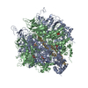
| ||||||||
| Unit cell |
|
-Components
-PROTEIN (F1-ATPASE ... , 3 types, 3 molecules ABG
| #1: Protein | Mass: 55348.219 Da / Num. of mol.: 1 / Fragment: ALPHA CHAIN / Source method: isolated from a natural source / Source: (natural) |
|---|---|
| #2: Protein | Mass: 51404.461 Da / Num. of mol.: 1 / Fragment: BETA CHAIN / Source method: isolated from a natural source / Source: (natural) |
| #3: Protein | Mass: 29974.396 Da / Num. of mol.: 1 / Fragment: GAMMA CHAIN / Source method: isolated from a natural source / Source: (natural) |
-Non-polymers , 5 types, 65 molecules 


| #4: Chemical | ChemComp-MG /  Mass: 24.305 Da / Num. of mol.: 1 / Source method: obtained synthetically / Formula: Mg Mass: 24.305 Da / Num. of mol.: 1 / Source method: obtained synthetically / Formula: Mg |
|---|---|
| #5: Chemical | ChemComp-ATP /  Mass: 507.181 Da / Num. of mol.: 1 / Source method: obtained synthetically / Formula: C10H16N5O13P3 / Comment: ATP, energy-carrying molecule*YM Mass: 507.181 Da / Num. of mol.: 1 / Source method: obtained synthetically / Formula: C10H16N5O13P3 / Comment: ATP, energy-carrying molecule*YM |
| #6: Chemical | ChemComp-PO4 /  Mass: 94.971 Da / Num. of mol.: 1 / Source method: obtained synthetically / Formula: PO4 Mass: 94.971 Da / Num. of mol.: 1 / Source method: obtained synthetically / Formula: PO4 |
| #7: Chemical | ChemComp-ADP /  Mass: 427.201 Da / Num. of mol.: 1 / Source method: obtained synthetically / Formula: C10H15N5O10P2 / Comment: ADP, energy-carrying molecule*YM Mass: 427.201 Da / Num. of mol.: 1 / Source method: obtained synthetically / Formula: C10H15N5O10P2 / Comment: ADP, energy-carrying molecule*YM |
| #8: Water | ChemComp-HOH / Mass: 18.015 Da / Num. of mol.: 61 / Source method: isolated from a natural source / Formula: H2O |
-Experimental details
-Experiment
| Experiment | Method: X-RAY DIFFRACTION / Number of used crystals: 1 |
|---|
- Sample preparation
| Crystal | Density Matthews: 2.62 Å3/Da / Density % sol: 50 % | |||||||||||||||||||||||||||||||||||
|---|---|---|---|---|---|---|---|---|---|---|---|---|---|---|---|---|---|---|---|---|---|---|---|---|---|---|---|---|---|---|---|---|---|---|---|---|
| Crystal grow | pH: 7.14 Details: 2.1 M AMMONIUM SULFATE AS PRECIPITANT, 0.2 M KPI (PH 7.14) BUFFER, 0.005 M ATP. AT ROOM TEMPERATURE Temp details: room temp | |||||||||||||||||||||||||||||||||||
| Crystal grow | *PLUS pH: 7.45 / Method: microdialysis / Details: Amzel, L.M., (1978) J.Biol.Chem., 253, 2067. | |||||||||||||||||||||||||||||||||||
| Components of the solutions | *PLUS
|
-Data collection
| Diffraction | Mean temperature: 100 K |
|---|---|
| Diffraction source | Source: ROTATING ANODE / Type: RIGAKU RU200 / Wavelength: 1.5418 |
| Detector | Type: MACSCIENCE / Detector: IMAGE PLATE / Details: MONOCROMATOR |
| Radiation | Monochromator: GRAPHITE / Protocol: SINGLE WAVELENGTH / Monochromatic (M) / Laue (L): M / Scattering type: x-ray |
| Radiation wavelength | Wavelength: 1.5418 Å / Relative weight: 1 |
| Reflection | Resolution: 2.8→100 Å / Num. obs: 35334 / % possible obs: 85.3 % / Redundancy: 11 % / Rsym value: 0.103 / Net I/σ(I): 7.1 |
| Reflection | *PLUS Rmerge(I) obs: 0.103 |
| Reflection shell | *PLUS Mean I/σ(I) obs: 7.1 |
- Processing
| Software | Name: X-PLOR / Version: 3.851 / Classification: refinement | ||||||||||||||||||||||||||||||||||||||||||||||||||||||||||||
|---|---|---|---|---|---|---|---|---|---|---|---|---|---|---|---|---|---|---|---|---|---|---|---|---|---|---|---|---|---|---|---|---|---|---|---|---|---|---|---|---|---|---|---|---|---|---|---|---|---|---|---|---|---|---|---|---|---|---|---|---|---|
| Refinement | Method to determine structure: MIR / Resolution: 2.8→6 Å / Cross valid method: THROUGHOUT / σ(F): 3 Details: FURTHER REFINEMENT IS IN PROGRESS. BECAUSE OF THE CRYSTAL SYMMETRY, THE INTERPRETABLE REGIONS OF THE GAMMA SUBUNIT ARE SEEN AS PARTIALLY OCCUPIED OVERLAPING COPIES. GAMMA AND SMALL REGIONS ...Details: FURTHER REFINEMENT IS IN PROGRESS. BECAUSE OF THE CRYSTAL SYMMETRY, THE INTERPRETABLE REGIONS OF THE GAMMA SUBUNIT ARE SEEN AS PARTIALLY OCCUPIED OVERLAPING COPIES. GAMMA AND SMALL REGIONS OF ALPHA AND BETA-SUBUNIT INTERACTING WITH GAMMA ARE REFINED WITH A PARTIAL OCUPPANCY.
| ||||||||||||||||||||||||||||||||||||||||||||||||||||||||||||
| Refinement step | Cycle: LAST / Resolution: 2.8→6 Å
| ||||||||||||||||||||||||||||||||||||||||||||||||||||||||||||
| Refine LS restraints |
| ||||||||||||||||||||||||||||||||||||||||||||||||||||||||||||
| Software | *PLUS Name: X-PLOR / Version: 3.851 / Classification: refinement | ||||||||||||||||||||||||||||||||||||||||||||||||||||||||||||
| Refinement | *PLUS Highest resolution: 2.8 Å / Lowest resolution: 6 Å / σ(F): 3 / % reflection Rfree: 2.5 % / Rfactor Rfree: 0.29 | ||||||||||||||||||||||||||||||||||||||||||||||||||||||||||||
| Solvent computation | *PLUS | ||||||||||||||||||||||||||||||||||||||||||||||||||||||||||||
| Displacement parameters | *PLUS | ||||||||||||||||||||||||||||||||||||||||||||||||||||||||||||
| Refine LS restraints | *PLUS
|
