 Movie
Movie Controller

[English] 日本語
 Yorodumi
- PDB-1m7v: STRUCTURE OF A NITRIC OXIDE SYNTHASE HEME PROTEIN FROM BACILLUS S... -
+ Open data

- Basic information
| Entry | Database: PDB / ID: 1m7v | ||||||
|---|---|---|---|---|---|---|---|
| Title | STRUCTURE OF A NITRIC OXIDE SYNTHASE HEME PROTEIN FROM BACILLUS SUBTILIS WITH TETRAHYDROFOLATE AND ARGININE BOUND | ||||||
 Components Components | Nitric Oxide Synthase | ||||||
 Keywords Keywords | OXIDOREDUCTASE / pterin oxygenase / bacteria / nitric oxide / heme | ||||||
| Function / homology |  Function and homology information Function and homology informationnitric-oxide synthase (flavodoxin) / nitric-oxide synthase activity / nitric oxide biosynthetic process / heme binding / metal ion binding Similarity search - Function | ||||||
| Biological species |  | ||||||
| Method |  X-RAY DIFFRACTION / SYNCHROTRON / MOLECULAR REPLACEMENT / Resolution: 1.95 Å X-RAY DIFFRACTION / SYNCHROTRON / MOLECULAR REPLACEMENT / Resolution: 1.95 Å | ||||||
 Authors Authors | Pant, K. / Bilwes, A.M. / Adak, S. / Stuehr, D.J. / Crane, B.R. | ||||||
 Citation Citation | Journal: Biochemistry / Year: 2002 Title: Structure of a nitric oxide synthase heme protein from Bacillus subtilis. Authors: Pant, K. / Bilwes, A.M. / Adak, S. / Stuehr, D.J. / Crane, B.R. | ||||||
| History |
| ||||||
| Remark 999 | SEQUENCE AT THE TIME OF PROCESSING, THIS SEQUENCE OF CHAIN A HAS NOT YET BEEN DEPOSITED IN A ...SEQUENCE AT THE TIME OF PROCESSING, THIS SEQUENCE OF CHAIN A HAS NOT YET BEEN DEPOSITED IN A SEQUENCE DATABASE. |
- Structure visualization
| Structure viewer | Molecule: MolmilJmol/JSmol |
|---|
- Downloads & links
-Download
| PDBx/mmCIF format | 1m7v.cif.gz | 96.5 KB | Display | PDBx/mmCIF format |
|---|---|---|---|---|
| PDB format | pdb1m7v.ent.gz | 71.2 KB | Display | PDB format |
| PDBx/mmJSON format | 1m7v.json.gz | Tree view | PDBx/mmJSON format | |
| Others |  Other downloads Other downloads |
-Validation report
| Arichive directory | https://data.pdbj.org/pub/pdb/validation_reports/m7/1m7vftp://data.pdbj.org/pub/pdb/validation_reports/m7/1m7v | HTTPS FTP |
|---|
-Related structure data
| Related structure data |  1m7zC  1dwwS S: Starting model for refinement C: citing same article ( |
|---|---|
| Similar structure data |


-Links
 PDBj
PDBj
- Assembly
| Deposited unit | 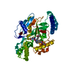
| ||||||||
|---|---|---|---|---|---|---|---|---|---|
| 1 | 
| ||||||||
| Unit cell |
| ||||||||
| Components on special symmetry positions |
| ||||||||
| Details | The second half of the biologically active dimer is generated by the crystallographic two fold axis: -x, -y, z |
-Components
| #1: Protein | Mass: 42097.227 Da / Num. of mol.: 1 Source method: isolated from a genetically manipulated source Source: (gene. exp.) |
|---|---|
| #2: Chemical | ChemComp-ARG /   Type: L-peptide linking / Mass: 175.209 Da / Num. of mol.: 1 / Source method: obtained synthetically / Formula: C6H15N4O2 Type: L-peptide linking / Mass: 175.209 Da / Num. of mol.: 1 / Source method: obtained synthetically / Formula: C6H15N4O2 |
| #3: Chemical | ChemComp-HEM /   Mass: 616.487 Da / Num. of mol.: 1 / Source method: obtained synthetically / Formula: C34H32FeN4O4 Mass: 616.487 Da / Num. of mol.: 1 / Source method: obtained synthetically / Formula: C34H32FeN4O4 |
| #4: Chemical | ChemComp-THG / (  Mass: 445.429 Da / Num. of mol.: 1 / Source method: obtained synthetically / Formula: C19H23N7O6 Mass: 445.429 Da / Num. of mol.: 1 / Source method: obtained synthetically / Formula: C19H23N7O6 |
| #5: Water | ChemComp-HOH /  Mass: 18.015 Da / Num. of mol.: 258 / Source method: isolated from a natural source / Formula: H2O Mass: 18.015 Da / Num. of mol.: 258 / Source method: isolated from a natural source / Formula: H2O |
| Has protein modification | N |
-Experimental details
-Experiment
| Experiment | Method: X-RAY DIFFRACTION / Number of used crystals: 1 |
|---|
- Sample preparation
| Crystal | Density Matthews: 2.81 Å3/Da / Density % sol: 56.17 % | |||||||||||||||||||||||||||||||||||||||||||||||||||||||||||||||
|---|---|---|---|---|---|---|---|---|---|---|---|---|---|---|---|---|---|---|---|---|---|---|---|---|---|---|---|---|---|---|---|---|---|---|---|---|---|---|---|---|---|---|---|---|---|---|---|---|---|---|---|---|---|---|---|---|---|---|---|---|---|---|---|---|
| Crystal grow | Temperature: 295 K / Method: vapor diffusion / pH: 7 Details: PEG 8K, potassium acetate, pH 7.0, VAPOR DIFFUSION, temperature 295K | |||||||||||||||||||||||||||||||||||||||||||||||||||||||||||||||
| Crystal grow | *PLUS Temperature: 22 ℃ / pH: 7.5 | |||||||||||||||||||||||||||||||||||||||||||||||||||||||||||||||
| Components of the solutions | *PLUS
|
-Data collection
| Diffraction | Mean temperature: 100 K |
|---|---|
| Diffraction source | Source: SYNCHROTRON / Site: CHESS  / Beamline: F1 / Wavelength: 0.916 Å / Beamline: F1 / Wavelength: 0.916 Å |
| Detector | Type: ADSC QUANTUM 4 / Detector: CCD / Date: May 24, 2002 |
| Radiation | Monochromator: Si / Protocol: SINGLE WAVELENGTH / Monochromatic (M) / Laue (L): M / Scattering type: x-ray |
| Radiation wavelength | Wavelength: 0.916 Å / Relative weight: 1 |
| Reflection | Resolution: 1.94→30 Å / Num. all: 44343 / Num. obs: 44343 / % possible obs: 99.7 % / Observed criterion σ(F): 0 / Observed criterion σ(I): -3 / Redundancy: 8.9 % / Biso Wilson estimate: 29.3 Å2 / Rsym value: 0.059 / Net I/σ(I): 27.5 |
| Reflection shell | Resolution: 1.94→1.98 Å / Mean I/σ(I) obs: 2.5 / Rsym value: 0.399 / % possible all: 99.7 |
| Reflection | *PLUS Num. measured all: 395408 / Rmerge(I) obs: 0.059 |
| Reflection shell | *PLUS % possible obs: 99.7 % / Rmerge(I) obs: 0.399 |
- Processing
| Software |
| ||||||||||||||||||||||||||||||||||||
|---|---|---|---|---|---|---|---|---|---|---|---|---|---|---|---|---|---|---|---|---|---|---|---|---|---|---|---|---|---|---|---|---|---|---|---|---|---|
| Refinement | Method to determine structure: MOLECULAR REPLACEMENT Starting model: PDB ENTRY 1DWW Resolution: 1.95→29.8 Å / Rfactor Rfree error: 0.006 / Data cutoff high absF: 1406462.31 / Data cutoff high rms absF: 1406462.31 / Data cutoff low absF: 0 / Isotropic thermal model: RESTRAINED / Cross valid method: THROUGHOUT / σ(F): 0 / Stereochemistry target values: Engh & Huber Details: The glutamate moiety of tetrahydrofolate is not well ordered and likely has alternate conformations
| ||||||||||||||||||||||||||||||||||||
| Solvent computation | Solvent model: FLAT MODEL / Bsol: 27.1565 Å2 / ksol: 0.338359 e/Å3 | ||||||||||||||||||||||||||||||||||||
| Displacement parameters | Biso mean: 42.7 Å2
| ||||||||||||||||||||||||||||||||||||
| Refine analyze |
| ||||||||||||||||||||||||||||||||||||
| Refinement step | Cycle: LAST / Resolution: 1.95→29.8 Å
| ||||||||||||||||||||||||||||||||||||
| Refine LS restraints |
| ||||||||||||||||||||||||||||||||||||
| LS refinement shell | Resolution: 1.95→2.07 Å / Rfactor Rfree error: 0.02 / Total num. of bins used: 6
| ||||||||||||||||||||||||||||||||||||
| Xplor file |
| ||||||||||||||||||||||||||||||||||||
| Refinement | *PLUS Highest resolution: 1.94 Å / % reflection Rfree: 10 % / Rfactor Rfree: 0.239 / Rfactor Rwork: 0.225 | ||||||||||||||||||||||||||||||||||||
| Solvent computation | *PLUS | ||||||||||||||||||||||||||||||||||||
| Displacement parameters | *PLUS | ||||||||||||||||||||||||||||||||||||
| Refine LS restraints | *PLUS
| ||||||||||||||||||||||||||||||||||||
| LS refinement shell | *PLUS Highest resolution: 1.94 Å / Lowest resolution: 1.98 Å / Rfactor Rfree: 0.333 / Rfactor Rwork: 0.322 |
