 Movie
Movie Controller

[English] 日本語
 Yorodumi
- PDB-1m0d: Crystal Structure of Bacteriophage T7 Endonuclease I with a Wild-... -
+ Open data

- Basic information
| Entry | Database: PDB / ID: 1m0d | ||||||
|---|---|---|---|---|---|---|---|
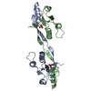
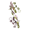
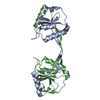
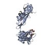
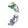
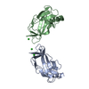
| Title | Crystal Structure of Bacteriophage T7 Endonuclease I with a Wild-Type Active Site and Bound Manganese Ions | ||||||
 Components Components | Endodeoxyribonuclease I | ||||||
 Keywords Keywords | HYDROLASE / Holliday junction resolvase / Homodimer / Domain Swapped / Composite active site | ||||||
| Function / homology |  Function and homology information Function and homology informationdegradation of host chromosome by virus / deoxyribonuclease IV / deoxyribonuclease IV (phage-T4-induced) activity / double-stranded DNA endonuclease activity / crossover junction DNA endonuclease activity / DNA integration / symbiont-mediated suppression of host gene expression / DNA binding Similarity search - Function | ||||||
| Biological species |   Enterobacteria phage T7 (virus) Enterobacteria phage T7 (virus) | ||||||
| Method |  X-RAY DIFFRACTION / SYNCHROTRON / MOLECULAR REPLACEMENT / Resolution: 1.9 Å X-RAY DIFFRACTION / SYNCHROTRON / MOLECULAR REPLACEMENT / Resolution: 1.9 Å | ||||||
 Authors Authors | Hadden, J.M. / Declais, A.C. / Phillips, S.E. / Lilley, D.M. | ||||||
 Citation Citation | Journal: EMBO J. / Year: 2002 Title: Metal ions bound at the active site of the junction-resolving enzyme T7 endonuclease I. Authors: Hadden, J.M. / Declais, A.C. / Phillips, S.E. / Lilley, D.M. | ||||||
| History |
|
- Structure visualization
| Structure viewer | Molecule: MolmilJmol/JSmol |
|---|
- Downloads & links
-Download
| PDBx/mmCIF format | 1m0d.cif.gz | 129 KB | Display | PDBx/mmCIF format |
|---|---|---|---|---|
| PDB format | pdb1m0d.ent.gz | 99.9 KB | Display | PDB format |
| PDBx/mmJSON format | 1m0d.json.gz | Tree view | PDBx/mmJSON format | |
| Others |  Other downloads Other downloads |
-Validation report
| Arichive directory | https://data.pdbj.org/pub/pdb/validation_reports/m0/1m0dftp://data.pdbj.org/pub/pdb/validation_reports/m0/1m0d | HTTPS FTP |
|---|
-Related structure data
| Related structure data |  1m0iC  1fzrS S: Starting model for refinement C: citing same article ( |
|---|---|
| Similar structure data |

-Links
 PDBj
PDBj


- Assembly
| Deposited unit | 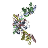
| ||||||||
|---|---|---|---|---|---|---|---|---|---|
| 1 | 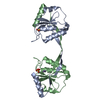
| ||||||||
| 2 | 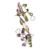
| ||||||||
| Unit cell |
| ||||||||
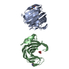
| Details | Endonuclease I is active as a homodimer. There are 2 homodimers in the asymmetric unit. Chains A and B form one homodimer. |
-Components
| #1: Protein | Mass: 16069.490 Da / Num. of mol.: 4 / Fragment: Residues 12-149 Source method: isolated from a genetically manipulated source Source: (gene. exp.) Enterobacteria phage T7 (virus) / Genus: T7-like viruses / Gene: Endonuclease I / Plasmid: pET 19B / Species (production host): Escherichia coli / Production host:  #2: Chemical | ChemComp-MN /   Mass: 54.938 Da / Num. of mol.: 8 / Source method: obtained synthetically / Formula: Mn Mass: 54.938 Da / Num. of mol.: 8 / Source method: obtained synthetically / Formula: Mn#3: Chemical | ChemComp-SO4 /   Mass: 96.063 Da / Num. of mol.: 4 / Source method: obtained synthetically / Formula: SO4 Mass: 96.063 Da / Num. of mol.: 4 / Source method: obtained synthetically / Formula: SO4#4: Water | ChemComp-HOH / |  Mass: 18.015 Da / Num. of mol.: 473 / Source method: isolated from a natural source / Formula: H2O Mass: 18.015 Da / Num. of mol.: 473 / Source method: isolated from a natural source / Formula: H2O |
|---|
-Experimental details
-Experiment
| Experiment | Method: X-RAY DIFFRACTION / Number of used crystals: 1 |
|---|
- Sample preparation
| Crystal | Density Matthews: 3.96 Å3/Da / Density % sol: 68.96 % | |||||||||||||||||||||||||||||||||||||||||||||||||
|---|---|---|---|---|---|---|---|---|---|---|---|---|---|---|---|---|---|---|---|---|---|---|---|---|---|---|---|---|---|---|---|---|---|---|---|---|---|---|---|---|---|---|---|---|---|---|---|---|---|---|
| Crystal grow | Temperature: 291 K Method: vapor diffusion, hanging drop. seeds of e65k mutant used pH: 7.2 Details: PEG 4000, Ammonium sulphate, Sodium chloride, Tris HCL, pH 7.2, Vapor diffusion, hanging drop. Seeds of E65K mutant used, temperature 291K | |||||||||||||||||||||||||||||||||||||||||||||||||
| Crystal grow | *PLUS pH: 8 / Method: vapor diffusion, hanging drop | |||||||||||||||||||||||||||||||||||||||||||||||||
| Components of the solutions | *PLUS
|
-Data collection
| Diffraction | Mean temperature: 100 K |
|---|---|
| Diffraction source | Source: SYNCHROTRON / Site: ESRF  / Beamline: ID14-2 / Wavelength: 0.933 Å / Beamline: ID14-2 / Wavelength: 0.933 Å |
| Detector | Type: ADSC QUANTUM 4 / Detector: CCD / Date: Jun 25, 2001 |
| Radiation | Monochromator: Diamond (111), Ge (220) / Protocol: SINGLE WAVELENGTH / Monochromatic (M) / Laue (L): M / Scattering type: x-ray |
| Radiation wavelength | Wavelength: 0.933 Å / Relative weight: 1 |
| Reflection | Resolution: 1.9→25.55 Å / Num. all: 81257 / Num. obs: 81257 / % possible obs: 99.9 % / Observed criterion σ(I): 0 / Redundancy: 5.2 % / Rsym value: 0.058 / Net I/σ(I): 7.6 |
| Reflection shell | Resolution: 1.9→1.99 Å / Redundancy: 4.4 % / Mean I/σ(I) obs: 1.6 / Num. unique all: 11678 / Rsym value: 0.36 / % possible all: 99.4 |
| Reflection | *PLUS Num. measured all: 419157 / Rmerge(I) obs: 0.085 |
| Reflection shell | *PLUS % possible obs: 99.4 % |
- Processing
| Software |
| ||||||||||||||||||||
|---|---|---|---|---|---|---|---|---|---|---|---|---|---|---|---|---|---|---|---|---|---|
| Refinement | Method to determine structure: MOLECULAR REPLACEMENT Starting model: PDB ENTRY 1fzr Resolution: 1.9→24.8 Å / Rfactor Rfree error: 0.004 / Data cutoff high absF: 1676632.53 / Data cutoff high rms absF: 1676632.53 / Data cutoff low absF: 0 / Isotropic thermal model: RESTRAINED / Cross valid method: THROUGHOUT / σ(F): 0 / Stereochemistry target values: Engh & Huber Details: Maximum Likelihood target used as implemented in CNS
| ||||||||||||||||||||
| Solvent computation | Solvent model: FLAT MODEL / Bsol: 59.3311 Å2 / ksol: 0.380933 e/Å3 | ||||||||||||||||||||
| Displacement parameters | Biso mean: 40.4 Å2
| ||||||||||||||||||||
| Refine analyze | Luzzati coordinate error free: 0.26 Å / Luzzati sigma a free: 0.15 Å | ||||||||||||||||||||
| Refinement step | Cycle: LAST / Resolution: 1.9→24.8 Å
| ||||||||||||||||||||
| Refine LS restraints |
| ||||||||||||||||||||
| LS refinement shell | Resolution: 1.9→1.97 Å / Rfactor Rfree error: 0.014 / Total num. of bins used: 10
| ||||||||||||||||||||
| Xplor file |
| ||||||||||||||||||||
| Refinement | *PLUS Highest resolution: 1.9 Å / % reflection Rfree: 5 % / Rfactor obs: 0.21 / Rfactor Rfree: 0.234 / Rfactor Rwork: 0.21 | ||||||||||||||||||||
| Solvent computation | *PLUS | ||||||||||||||||||||
| Displacement parameters | *PLUS | ||||||||||||||||||||
| Refine LS restraints | *PLUS
| ||||||||||||||||||||
| LS refinement shell | *PLUS Rfactor Rfree: 0.285 / Rfactor Rwork: 0.259 |
