 Movie
Movie Controller

[English] 日本語
 Yorodumi
- PDB-1ltr: CRYSTAL STRUCTURE OF THE B SUBUNIT OF HUMAN HEAT-LABILE ENTEROTOX... -
+ Open data

- Basic information
| Entry | Database: PDB / ID: 1ltr | ||||||
|---|---|---|---|---|---|---|---|
| Title | CRYSTAL STRUCTURE OF THE B SUBUNIT OF HUMAN HEAT-LABILE ENTEROTOXIN FROM E. COLI CARRYING A PEPTIDE WITH ANTI-HSV ACTIVITY | ||||||
 Components Components | HEAT-LABILE ENTEROTOXIN | ||||||
 Keywords Keywords | ENTEROTOXIN / B SUBUNIT / HEAT-LABILE ENTEROTOXIN / ANTI-HSV | ||||||
| Function / homology |  Function and homology information Function and homology information | ||||||
| Biological species |  | ||||||
| Method |  X-RAY DIFFRACTION / SYNCHROTRON / MOLECULAR REPLACEMENT / Resolution: 3.04 Å X-RAY DIFFRACTION / SYNCHROTRON / MOLECULAR REPLACEMENT / Resolution: 3.04 Å | ||||||
 Authors Authors | Matkovic-Calogovic, D. / Loreggian, A. / Palu, G. / Zanotti, G. | ||||||
 Citation Citation | Journal: J.Biol.Chem. / Year: 1999 Title: Crystal structure of the B subunit of Escherichia coli heat-labile enterotoxin carrying peptides with anti-herpes simplex virus type 1 activity. Authors: Matkovic-Calogovic, D. / Loregian, A. / D'Acunto, M.R. / Battistutta, R. / Tossi, A. / Palu, G. / Zanotti, G. #1: Journal: Proc.Natl.Acad.Sci.USA / Year: 1994Title: Specific Inhibition of Herpes Virus Replication by Receptor-Mediated Entry of an Antiviral Peptide Linked to Escherichia Coli Enterotoxin B Subunit Authors: Marcello, A. / Loregian, A. / Cross, A. / Marsden, H. / Hirst, T.R. / Palu, G. #2: Journal: J.Mol.Biol. / Year: 1993Title: Refined Structure of Escherichia Coli Heat-Labile Enterotoxin, a Close Relative of Cholera Toxin Authors: Sixma, T.K. / Kalk, K.H. / Van Zanten, B.A. / Dauter, Z. / Kingma, J. / Witholt, B. / Hol, W.G. | ||||||
| History |
|
- Structure visualization
| Structure viewer | Molecule: MolmilJmol/JSmol |
|---|
- Downloads & links
-Download
| PDBx/mmCIF format | 1ltr.cif.gz | 118.4 KB | Display | PDBx/mmCIF format |
|---|---|---|---|---|
| PDB format | pdb1ltr.ent.gz | 93.7 KB | Display | PDB format |
| PDBx/mmJSON format | 1ltr.json.gz | Tree view | PDBx/mmJSON format | |
| Others |  Other downloads Other downloads |
-Validation report
| Arichive directory | https://data.pdbj.org/pub/pdb/validation_reports/lt/1ltrftp://data.pdbj.org/pub/pdb/validation_reports/lt/1ltr | HTTPS FTP |
|---|
-Related structure data
| Related structure data |  1b44C 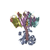 1ltsS S: Starting model for refinement C: citing same article ( |
|---|---|
| Similar structure data |


-Links
 PDBj
PDBj
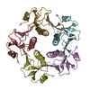
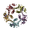
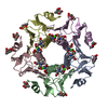
- Assembly
| Deposited unit | 
| |||||||||||||||||||||||||||||||||||
|---|---|---|---|---|---|---|---|---|---|---|---|---|---|---|---|---|---|---|---|---|---|---|---|---|---|---|---|---|---|---|---|---|---|---|---|---|
| 1 |
| |||||||||||||||||||||||||||||||||||
| 2 | 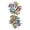
| |||||||||||||||||||||||||||||||||||
| Unit cell |
| |||||||||||||||||||||||||||||||||||
| Noncrystallographic symmetry (NCS) | NCS domain:
NCS oper:
|
-Components
| #1: Protein | Mass: 12747.591 Da / Num. of mol.: 5 / Fragment: SUBUNIT B-R2 / Mutation: N103K Source method: isolated from a genetically manipulated source Details: THE B SUBUNIT HAS A PEPTIDE WITH ANTI-HSV ACTIVITY AS AN EXTENSION, RESIDUES 104-113 Source: (gene. exp.) Vibrio sp. (bacteria) / Strain (production host): SP60 / References: UniProt: P13811, UniProt: P0CK94*PLUS#2: Chemical | ChemComp-SO4 /   Mass: 96.063 Da / Num. of mol.: 6 / Source method: obtained synthetically / Formula: SO4 Mass: 96.063 Da / Num. of mol.: 6 / Source method: obtained synthetically / Formula: SO4#3: Water | ChemComp-HOH / |  Mass: 18.015 Da / Num. of mol.: 116 / Source method: isolated from a natural source / Formula: H2O Mass: 18.015 Da / Num. of mol.: 116 / Source method: isolated from a natural source / Formula: H2OHas protein modification | Y | |
|---|
-Experimental details
-Experiment
| Experiment | Method: X-RAY DIFFRACTION / Number of used crystals: 1 |
|---|
- Sample preparation
| Crystal | Density Matthews: 5.54 Å3/Da / Density % sol: 78 % | ||||||||||||||||||||||||||||||
|---|---|---|---|---|---|---|---|---|---|---|---|---|---|---|---|---|---|---|---|---|---|---|---|---|---|---|---|---|---|---|---|
| Crystal grow | pH: 6 / Details: pH 6 | ||||||||||||||||||||||||||||||
| Crystal | *PLUS Density % sol: 77 % | ||||||||||||||||||||||||||||||
| Crystal grow | *PLUS Temperature: 4 ℃ / Method: vapor diffusion, hanging drop | ||||||||||||||||||||||||||||||
| Components of the solutions | *PLUS
|
-Data collection
| Diffraction | Mean temperature: 100 K |
|---|---|
| Diffraction source | Source: SYNCHROTRON / Site: ELETTRA  / Beamline: 5.2R / Wavelength: 1.4 / Beamline: 5.2R / Wavelength: 1.4 |
| Detector | Type: MARRESEARCH / Detector: IMAGE PLATE AREA DETECTOR / Date: Feb 1, 1998 / Details: MIRRORS |
| Radiation | Monochromator: SI(111) / Monochromatic (M) / Laue (L): M / Scattering type: x-ray |
| Radiation wavelength | Wavelength: 1.4 Å / Relative weight: 1 |
| Reflection | Resolution: 3.04→23.19 Å / Num. obs: 27477 / % possible obs: 97.5 % / Observed criterion σ(I): 2 / Redundancy: 3.5 % / Biso Wilson estimate: 40 Å2 / Rmerge(I) obs: 0.041 / Rsym value: 0.044 / Net I/σ(I): 11.8 |
| Reflection shell | Resolution: 3.04→3.39 Å / Redundancy: 3.4 % / Rmerge(I) obs: 0.073 / Mean I/σ(I) obs: 0.1 / % possible all: 95.6 |
| Reflection | *PLUS Highest resolution: 3 Å / Lowest resolution: 23.2 Å / Num. measured all: 97277 |
- Processing
| Software |
| ||||||||||||||||||||||||||||||||||||||||||||||||||||||||||||
|---|---|---|---|---|---|---|---|---|---|---|---|---|---|---|---|---|---|---|---|---|---|---|---|---|---|---|---|---|---|---|---|---|---|---|---|---|---|---|---|---|---|---|---|---|---|---|---|---|---|---|---|---|---|---|---|---|---|---|---|---|---|
| Refinement | Method to determine structure: MOLECULAR REPLACEMENT Starting model: PDB ENTRY 1LTS, CHAINS D-H Resolution: 3.04→8 Å / Rfactor Rfree error: 0.005 / Data cutoff high absF: 1000000 / Data cutoff low absF: 0.001 / Isotropic thermal model: RESTRAINED / Cross valid method: THROUGHOUT / σ(F): 2
| ||||||||||||||||||||||||||||||||||||||||||||||||||||||||||||
| Displacement parameters | Biso mean: 23 Å2 | ||||||||||||||||||||||||||||||||||||||||||||||||||||||||||||
| Refine analyze | Luzzati d res low obs: 8 Å | ||||||||||||||||||||||||||||||||||||||||||||||||||||||||||||
| Refinement step | Cycle: LAST / Resolution: 3.04→8 Å
| ||||||||||||||||||||||||||||||||||||||||||||||||||||||||||||
| Refine LS restraints |
| ||||||||||||||||||||||||||||||||||||||||||||||||||||||||||||
| Refine LS restraints NCS | Refine-ID: X-RAY DIFFRACTION / Rms dev Biso : _ / Rms dev position: _ / Weight Biso : _ / Weight position: _
| ||||||||||||||||||||||||||||||||||||||||||||||||||||||||||||
| LS refinement shell | Resolution: 3.04→3.17 Å / Rfactor Rfree error: 0.02 / Total num. of bins used: 8
| ||||||||||||||||||||||||||||||||||||||||||||||||||||||||||||
| Xplor file |
| ||||||||||||||||||||||||||||||||||||||||||||||||||||||||||||
| Software | *PLUS Name: X-PLOR / Version: 3.84 / Classification: refinement | ||||||||||||||||||||||||||||||||||||||||||||||||||||||||||||
| Refinement | *PLUS Rfactor obs: 0.183 | ||||||||||||||||||||||||||||||||||||||||||||||||||||||||||||
| Solvent computation | *PLUS | ||||||||||||||||||||||||||||||||||||||||||||||||||||||||||||
| Displacement parameters | *PLUS | ||||||||||||||||||||||||||||||||||||||||||||||||||||||||||||
| Refine LS restraints | *PLUS
| ||||||||||||||||||||||||||||||||||||||||||||||||||||||||||||
| LS refinement shell | *PLUS Rfactor obs: 0.2786 |
