 Movie
Movie Controller

[English] 日本語
 Yorodumi
- PDB-1ltd: THE 2.6 ANGSTROMS REFINED STRUCTURE OF THE ESCHERICHIA COLI RECOM... -
+ Open data

- Basic information
| Entry | Database: PDB / ID: 1ltd | ||||||
|---|---|---|---|---|---|---|---|
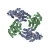
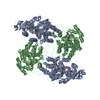
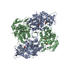
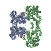
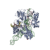
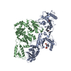
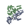
| Title | THE 2.6 ANGSTROMS REFINED STRUCTURE OF THE ESCHERICHIA COLI RECOMBINANT SACCHAROMYCES CEREVISIAE FLAVOCYTOCHROME B2-SULPHITE COMPLEX | ||||||
 Components Components | FLAVOCYTOCHROME B2 | ||||||
 Keywords Keywords | OXIDOREDUCTASE(CH-OH(D)-CYTOCHROME(A)) | ||||||
| Function / homology |  Function and homology information Function and homology informationL-lactate dehydrogenase (cytochrome) / L-lactate dehydrogenase (cytochrome) activity / lactate metabolic process / mitochondrial intermembrane space / mitochondrial inner membrane / heme binding / mitochondrion / metal ion binding / nucleus / cytosol Similarity search - Function | ||||||
| Biological species |  | ||||||
| Method |  X-RAY DIFFRACTION / Resolution: 2.6 Å X-RAY DIFFRACTION / Resolution: 2.6 Å | ||||||
 Authors Authors | Tegoni, M. / Cambillau, C. | ||||||
 Citation Citation | Journal: Protein Sci. / Year: 1994 Title: The 2.6-A refined structure of the Escherichia coli recombinant Saccharomyces cerevisiae flavocytochrome b2-sulfite complex. Authors: Tegoni, M. / Cambillau, C. #1: Journal: J.Mol.Biol. / Year: 1990Title: Molecular Structure of Flavocytochrome B2 at 2.4 Angstroms Resolution Authors: Xia, Z. / Mathews, F.S. #2: Journal: Biochem.J. / Year: 1989Title: High Level Expression of Fully Active Yeast Flavocytochrome B2 in Escherichia Coli Authors: Black, M. / White, S. / Reid, G. / Chapman, S. | ||||||
| History |
|
- Structure visualization
| Structure viewer | Molecule: MolmilJmol/JSmol |
|---|
- Downloads & links
-Download
| PDBx/mmCIF format | 1ltd.cif.gz | 191.2 KB | Display | PDBx/mmCIF format |
|---|---|---|---|---|
| PDB format | pdb1ltd.ent.gz | 149.4 KB | Display | PDB format |
| PDBx/mmJSON format | 1ltd.json.gz | Tree view | PDBx/mmJSON format | |
| Others |  Other downloads Other downloads |
-Validation report
| Arichive directory | https://data.pdbj.org/pub/pdb/validation_reports/lt/1ltdftp://data.pdbj.org/pub/pdb/validation_reports/lt/1ltd | HTTPS FTP |
|---|
-Related structure data
| Similar structure data |
|---|


-Links
 PDBj
PDBj


- Assembly
| Deposited unit | 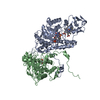
| ||||||||
|---|---|---|---|---|---|---|---|---|---|
| 1 | 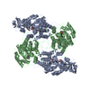
| ||||||||
| Unit cell |
| ||||||||
| Noncrystallographic symmetry (NCS) | NCS oper: (Code: given Matrix: (0.247, 0.4353, -0.8657), Vector: Details | THE ASYMMETRIC UNIT CONTAINS TWO SUBUNITS WHICH HAVE BEEN ASSIGNED CHAIN INDICATORS *A* AND *B*. THE TRANSFORMATION PRESENTED ON *MTRIX* RECORDS BELOW WILL YIELD APPROXIMATE COORDINATES FOR CHAIN *A* WHEN APPLIED TO CHAIN *B*. | |
-Components
| #1: Protein | Mass: 56080.242 Da / Num. of mol.: 2 Source method: isolated from a genetically manipulated source Source: (gene. exp.) References: UniProt: P00175, L-lactate dehydrogenase (cytochrome) #2: Chemical |   Mass: 80.063 Da / Num. of mol.: 2 / Source method: obtained synthetically / Formula: SO3 Mass: 80.063 Da / Num. of mol.: 2 / Source method: obtained synthetically / Formula: SO3#3: Chemical |   Mass: 456.344 Da / Num. of mol.: 2 / Source method: obtained synthetically / Formula: C17H21N4O9P Mass: 456.344 Da / Num. of mol.: 2 / Source method: obtained synthetically / Formula: C17H21N4O9P#4: Chemical | ChemComp-HEM / |   Mass: 616.487 Da / Num. of mol.: 1 / Source method: obtained synthetically / Formula: C34H32FeN4O4 Mass: 616.487 Da / Num. of mol.: 1 / Source method: obtained synthetically / Formula: C34H32FeN4O4#5: Water | ChemComp-HOH / |  Mass: 18.015 Da / Num. of mol.: 206 / Source method: isolated from a natural source / Formula: H2O Mass: 18.015 Da / Num. of mol.: 206 / Source method: isolated from a natural source / Formula: H2OCompound details | EACH SUBUNIT CONTAINS TWO DOMAINS, A CYTOCHROME DOMAIN COMPRISING RESIDUES 5 - 99 AND A FLAVIN- ...EACH SUBUNIT CONTAINS TWO DOMAINS, A CYTOCHROME | |
|---|
-Experimental details
-Experiment
| Experiment | Method: X-RAY DIFFRACTION |
|---|
- Sample preparation
| Crystal | Density Matthews: 3.97 Å3/Da / Density % sol: 69 % | ||||||||||||||||||||||||||||||
|---|---|---|---|---|---|---|---|---|---|---|---|---|---|---|---|---|---|---|---|---|---|---|---|---|---|---|---|---|---|---|---|
| Crystal grow | *PLUS Temperature: 20 ℃ / pH: 6.5 / Method: vapor diffusion, sitting drop | ||||||||||||||||||||||||||||||
| Components of the solutions | *PLUS
|
-Data collection
| Radiation | Scattering type: x-ray |
|---|---|
| Radiation wavelength | Relative weight: 1 |
| Reflection | *PLUS Highest resolution: 2.6 Å / Lowest resolution: 100 Å / Num. obs: 44282 / % possible obs: 81 % / Num. measured all: 452385 / Rmerge(I) obs: 0.147 |
| Reflection shell | *PLUS Highest resolution: 2.6 Å / Lowest resolution: 2.9 Å / % possible obs: 55 % |
- Processing
| Software |
| ||||||||||||||||||||||||||||||||||||||||||||||||||||||||||||
|---|---|---|---|---|---|---|---|---|---|---|---|---|---|---|---|---|---|---|---|---|---|---|---|---|---|---|---|---|---|---|---|---|---|---|---|---|---|---|---|---|---|---|---|---|---|---|---|---|---|---|---|---|---|---|---|---|---|---|---|---|---|
| Refinement | Resolution: 2.6→6 Å / Rfactor Rwork: 0.173 / Rfactor obs: 0.173 / σ(F): 1 | ||||||||||||||||||||||||||||||||||||||||||||||||||||||||||||
| Refinement step | Cycle: LAST / Resolution: 2.6→6 Å
| ||||||||||||||||||||||||||||||||||||||||||||||||||||||||||||
| Refine LS restraints |
| ||||||||||||||||||||||||||||||||||||||||||||||||||||||||||||
| Software | *PLUS Name: X-PLOR / Classification: refinement | ||||||||||||||||||||||||||||||||||||||||||||||||||||||||||||
| Refinement | *PLUS Rfactor obs: 0.173 | ||||||||||||||||||||||||||||||||||||||||||||||||||||||||||||
| Solvent computation | *PLUS | ||||||||||||||||||||||||||||||||||||||||||||||||||||||||||||
| Displacement parameters | *PLUS | ||||||||||||||||||||||||||||||||||||||||||||||||||||||||||||
| Refine LS restraints | *PLUS Type: x_angle_d / Dev ideal: 3.37 |
