 Movie
Movie Controller


+ Open data

- Basic information
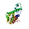
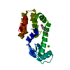
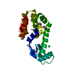
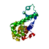
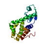
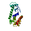
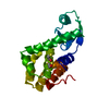
| Entry | Database: PDB / ID: 1lpy | ||||||
|---|---|---|---|---|---|---|---|
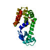
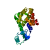
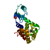
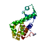
| Title | Multiple Methionine Substitutions in T4 Lysozyme | ||||||
 Components Components | LYSOZYME | ||||||
 Keywords Keywords | HYDROLASE / hydrolase (o-glycosyl) / T4 lysozyme / methionine core mutant / protein engineering / protein folding | ||||||
| Function / homology |  Function and homology information Function and homology informationviral release from host cell by cytolysis / peptidoglycan catabolic process / cell wall macromolecule catabolic process / lysozyme / lysozyme activity / host cell cytoplasm / defense response to bacterium Similarity search - Function | ||||||
| Biological species |  Enterobacteria phage T4 (virus) Enterobacteria phage T4 (virus) | ||||||
| Method |  X-RAY DIFFRACTION / MOLECULAR REPLACEMENT / Resolution: 1.65 Å X-RAY DIFFRACTION / MOLECULAR REPLACEMENT / Resolution: 1.65 Å | ||||||
 Authors Authors | Gassner, N.C. / Baase, W.A. / Mooers, B.H.M. / Busam, R.D. / Weaver, L.H. / Lindstrom, J.D. / Quillin, M.L. / Matthews, B.W. | ||||||
 Citation Citation | Journal: Biophys.Chem. / Year: 2003 Title: Multiple methionine substitutions are tolerated in T4 lysozyme and have coupled effects on folding and stability. Authors: Gassner, N.C. / Baase, W.A. / Mooers, B.H. / Busam, R.D. / Weaver, L.H. / Lindstrom, J.D. / Quillin, M.L. / Matthews, B.W. | ||||||
| History |
|
- Structure visualization
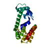
| Structure viewer | Molecule: MolmilJmol/JSmol |
|---|
- Downloads & links
-Download
| PDBx/mmCIF format | 1lpy.cif.gz | 49.3 KB | Display | PDBx/mmCIF format |
|---|---|---|---|---|
| PDB format | pdb1lpy.ent.gz | 35.5 KB | Display | PDB format |
| PDBx/mmJSON format | 1lpy.json.gz | Tree view | PDBx/mmJSON format | |
| Others |  Other downloads Other downloads |
-Validation report
| Arichive directory | https://data.pdbj.org/pub/pdb/validation_reports/lp/1lpyftp://data.pdbj.org/pub/pdb/validation_reports/lp/1lpy | HTTPS FTP |
|---|
-Related structure data
| Related structure data | 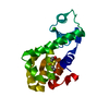 1ks3C 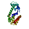 1kw5C  1kw7C 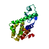 1ky0C 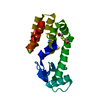 1ky1C 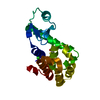 1l0jC 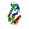 1l0kC 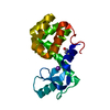 1lw9C 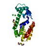 1lwgC 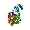 1lwkC C: citing same article ( |
|---|---|
| Similar structure data |


-Links
 PDBj
PDBj


- Assembly
| Deposited unit | 
| ||||||||
|---|---|---|---|---|---|---|---|---|---|
| 1 |
| ||||||||
| Unit cell |
|
-Components
| #1: Protein | Mass: 19654.395 Da / Num. of mol.: 1 Mutation: C54T,L84M,V87M,L91M,C97A,L99M,I100M,V103M,G110R,V111M,L118M,L121M,L133M Source method: isolated from a genetically manipulated source Source: (gene. exp.) Enterobacteria phage T4 (virus) / Genus: T4-like viruses / Species: Enterobacteria phage T4 sensu lato / Gene: E / Plasmid: phs1403 / Production host:  |
|---|---|
| #2: Chemical | ChemComp-PO4 /   Mass: 94.971 Da / Num. of mol.: 1 / Source method: obtained synthetically / Formula: PO4 Mass: 94.971 Da / Num. of mol.: 1 / Source method: obtained synthetically / Formula: PO4 |
| #3: Chemical | ChemComp-CL /   Mass: 35.453 Da / Num. of mol.: 1 / Source method: obtained synthetically / Formula: Cl Mass: 35.453 Da / Num. of mol.: 1 / Source method: obtained synthetically / Formula: Cl |
| #4: Chemical | ChemComp-BME /   Mass: 78.133 Da / Num. of mol.: 1 / Source method: obtained synthetically / Formula: C2H6OS Mass: 78.133 Da / Num. of mol.: 1 / Source method: obtained synthetically / Formula: C2H6OS |
| #5: Water | ChemComp-HOH /  Mass: 18.015 Da / Num. of mol.: 107 / Source method: isolated from a natural source / Formula: H2O Mass: 18.015 Da / Num. of mol.: 107 / Source method: isolated from a natural source / Formula: H2O |
-Experimental details
-Experiment
| Experiment | Method: X-RAY DIFFRACTION / Number of used crystals: 1 |
|---|
- Sample preparation
| Crystal | Density Matthews: 2.48 Å3/Da / Density % sol: 50.46 % | ||||||||||||||||||||||||||||||
|---|---|---|---|---|---|---|---|---|---|---|---|---|---|---|---|---|---|---|---|---|---|---|---|---|---|---|---|---|---|---|---|
| Crystal grow | *PLUS Method: vapor diffusion / Details: Eriksson, A.E., (1993) J. Mol. Biol., 229, 747. / PH range low: 7.1 / PH range high: 6.3 | ||||||||||||||||||||||||||||||
| Components of the solutions | *PLUS
|
-Data collection
| Radiation | Protocol: SINGLE WAVELENGTH / Monochromatic (M) / Laue (L): M / Scattering type: x-ray |
|---|---|
| Radiation wavelength | Relative weight: 1 |
| Reflection | Resolution: 1.65→15 Å / Num. all: 23682 / Num. obs: 23682 / % possible obs: 95 % / Observed criterion σ(I): 0 / Rmerge(I) obs: 0.043 / Net I/σ(I): 7.1 |
| Reflection shell | Resolution: 1.65→1.74 Å / Rmerge(I) obs: 0.103 / Mean I/σ(I) obs: 6.4 / Num. unique all: 1878 |
| Reflection | *PLUS Highest resolution: 1.7 Å / Lowest resolution: 15 Å / % possible obs: 96 % |
- Processing
| Software |
| ||||||||||||||||||
|---|---|---|---|---|---|---|---|---|---|---|---|---|---|---|---|---|---|---|---|
| Refinement | Method to determine structure: MOLECULAR REPLACEMENT Starting model: Native T4 Lysozyme Resolution: 1.65→15 Å / σ(F): 0 / Stereochemistry target values: TNT Details: Residues ASN 163 and LEU 164 are missing in the electron density.
| ||||||||||||||||||
| Refinement step | Cycle: LAST / Resolution: 1.65→15 Å
| ||||||||||||||||||
| Refine LS restraints |
| ||||||||||||||||||
| Refinement | *PLUS Highest resolution: 1.7 Å / Lowest resolution: 15 Å / Rfactor all: 0.209 / Rfactor obs: 0.207 | ||||||||||||||||||
| Solvent computation | *PLUS | ||||||||||||||||||
| Displacement parameters | *PLUS | ||||||||||||||||||
| Refine LS restraints | *PLUS Type: t_bond_d / Dev ideal: 0.017 |
