 Movie
Movie Controller

[English] 日本語
 Yorodumi
- PDB-1lhz: Structure of a Human Bence-Jones Dimer Crystallized in U.S. Space... -
+ Open data

- Basic information
| Entry | Database: PDB / ID: 1lhz | |||||||||
|---|---|---|---|---|---|---|---|---|---|---|
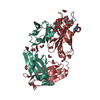
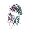
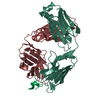
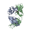
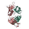
| Title | Structure of a Human Bence-Jones Dimer Crystallized in U.S. Space Shuttle Mission STS-95: 293K | |||||||||
 Components Components | IMMUNOGLOBULIN LAMBDA LIGHT CHAIN | |||||||||
 Keywords Keywords | IMMUNE SYSTEM / Human Bence-Jones Dimer / Microgravity Crystallization / Induced fit | |||||||||
| Function / homology |  Function and homology information Function and homology informationimmunoglobulin complex / adaptive immune response / extracellular region / plasma membrane Similarity search - Function | |||||||||
| Biological species |  Homo sapiens (human) Homo sapiens (human) | |||||||||
| Method |  X-RAY DIFFRACTION / MOLECULAR REPLACEMENT / Resolution: 2.3 Å X-RAY DIFFRACTION / MOLECULAR REPLACEMENT / Resolution: 2.3 Å | |||||||||
 Authors Authors | Terzyan, S.S. / DeWitt, C.R. / Ramsland, P.A. / Bourne, P.C. / Edmundson, A.B. | |||||||||
 Citation Citation | Journal: J.MOL.RECOG. / Year: 2003 Title: Comparison of the three-dimensional structures of a human Bence-Jones dimer crystallized on Earth and aboard US Space Shuttle Mission STS-95 Authors: Terzyan, S.S. / DeWitt, C.R. / Ramsland, P.A. / Bourne, P.C. / Edmundson, A.B. #1: Journal: Acta Crystallogr.,Sect.D / Year: 2002Title: Three-dimensional structure of an immunoglobulin light-chain dimer with amyloidogenic properties. Authors: BOURNE, P.C. / RAMSLAND, P.A. / SHAN, L. / Fan, Z.C. / DeWitt, C.R. / SHULTZ, B.B. / Terzyans, S.S. / Moomaw, C.R. / Slaughter, C.A. / Guddat, L.W. / Edmundson, A.B. | |||||||||
| History |
| |||||||||
| Remark 999 | sequence an appropriate sequence database reference was not available at the time of processing. |
- Structure visualization
| Structure viewer | Molecule: MolmilJmol/JSmol |
|---|
- Downloads & links
-Download
| PDBx/mmCIF format | 1lhz.cif.gz | 92.5 KB | Display | PDBx/mmCIF format |
|---|---|---|---|---|
| PDB format | pdb1lhz.ent.gz | 71.7 KB | Display | PDB format |
| PDBx/mmJSON format | 1lhz.json.gz | Tree view | PDBx/mmJSON format | |
| Others |  Other downloads Other downloads |
-Validation report
| Arichive directory | https://data.pdbj.org/pub/pdb/validation_reports/lh/1lhzftp://data.pdbj.org/pub/pdb/validation_reports/lh/1lhz | HTTPS FTP |
|---|
-Related structure data
| Related structure data |  1lgvC  1jvkS S: Starting model for refinement C: citing same article ( |
|---|---|
| Similar structure data |


-Links
 PDBj
PDBj

- Assembly
| Deposited unit | 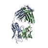
| ||||||||
|---|---|---|---|---|---|---|---|---|---|
| 1 |
| ||||||||
| Unit cell |
|
-Components
| #1: Antibody | Mass: 22608.980 Da / Num. of mol.: 2 / Source method: isolated from a natural source / Source: (natural) Homo sapiens (human) / References: UniProt: Q6PJG0*PLUS#2: Water | ChemComp-HOH / |  Mass: 18.015 Da / Num. of mol.: 168 / Source method: isolated from a natural source / Formula: H2O Mass: 18.015 Da / Num. of mol.: 168 / Source method: isolated from a natural source / Formula: H2OHas protein modification | Y | |
|---|
-Experimental details
-Experiment
| Experiment | Method: X-RAY DIFFRACTION / Number of used crystals: 1 |
|---|
- Sample preparation
| Crystal | Density Matthews: 2.63 Å3/Da / Density % sol: 43 % |
|---|---|
| Crystal grow | Temperature: 279 K / Method: vapor diffusion / Details: PEG 8000, VAPOR DIFFUSION, temperature 279K |
| Crystal grow | *PLUS Method: unknownDetails: Alvarado, U.R., (2001) J. Crystal Growth, 223, 407. |
-Data collection
| Diffraction | Mean temperature: 293 K |
|---|---|
| Diffraction source | Source: ROTATING ANODE / Type: RIGAKU RU300 / Wavelength: 1.54 Å |
| Detector | Type: MARRESEARCH / Detector: IMAGE PLATE / Date: Nov 19, 1998 / Details: Osmic multilayer |
| Radiation | Protocol: SINGLE WAVELENGTH / Monochromatic (M) / Laue (L): M / Scattering type: x-ray |
| Radiation wavelength | Wavelength: 1.54 Å / Relative weight: 1 |
| Reflection | Resolution: 2.3→25 Å / Num. obs: 21524 / % possible obs: 98.3 % / Observed criterion σ(I): -3 / Redundancy: 4.4 % / Biso Wilson estimate: 34.9 Å2 / Rmerge(I) obs: 0.077 / Net I/σ(I): 16.1 |
| Reflection shell | Resolution: 2.3→2.38 Å / Redundancy: 4.4 % / Rmerge(I) obs: 0.16 / Mean I/σ(I) obs: 7.6 / Num. unique all: 1943 / % possible all: 91.7 |
| Reflection | *PLUS Highest resolution: 2.3 Å / Num. obs: 21254 / Num. measured all: 92994 |
| Reflection shell | *PLUS Highest resolution: 2.3 Å / % possible obs: 91.7 % / Redundancy: 3 % / Num. unique obs: 1943 / Rmerge(I) obs: 0.161 |
- Processing
| Software |
| ||||||||||||||||||||||||||||
|---|---|---|---|---|---|---|---|---|---|---|---|---|---|---|---|---|---|---|---|---|---|---|---|---|---|---|---|---|---|
| Refinement | Method to determine structure: MOLECULAR REPLACEMENT Starting model: PDB ENTRY 1JVK Resolution: 2.3→25 Å / Cross valid method: R free / σ(F): 0 / Stereochemistry target values: Engh & Huber / Details: Used maximum likelihood target for amplitudes
| ||||||||||||||||||||||||||||
| Solvent computation | Bsol: 40.28 Å2 / ksol: 0.28 e/Å3 | ||||||||||||||||||||||||||||
| Displacement parameters | Biso mean: 34.85 Å2
| ||||||||||||||||||||||||||||
| Refinement step | Cycle: LAST / Resolution: 2.3→25 Å
| ||||||||||||||||||||||||||||
| Refine LS restraints |
| ||||||||||||||||||||||||||||
| Refinement | *PLUS Highest resolution: 2.3 Å | ||||||||||||||||||||||||||||
| Solvent computation | *PLUS | ||||||||||||||||||||||||||||
| Displacement parameters | *PLUS |
