 Movie
Movie Controller


+ Open data

- Basic information
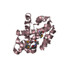
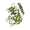
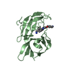
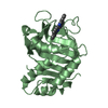
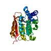
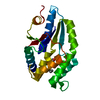
| Entry | Database: PDB / ID: 1lg7 | ||||||
|---|---|---|---|---|---|---|---|
| Title | Crystal structure of Vesicular Stomatitis Virus Matrix Protein | ||||||
 Components Components | VSV matrix protein | ||||||
 Keywords Keywords | VIRAL PROTEIN / VIRUS MATRIX | ||||||
| Function / homology |  Function and homology information Function and homology informationsymbiont-mediated suppression of host transcription initiation from RNA polymerase II promoter / host cell nuclear membrane / viral budding via host ESCRT complex / symbiont-mediated suppression of host mRNA export from nucleus / structural constituent of virion / host cell cytoplasm / viral envelope Similarity search - Function | ||||||
| Biological species |  Vesicular stomatitis virus Vesicular stomatitis virus | ||||||
| Method |  X-RAY DIFFRACTION / SYNCHROTRON / SIRAS / Resolution: 1.96 Å X-RAY DIFFRACTION / SYNCHROTRON / SIRAS / Resolution: 1.96 Å | ||||||
 Authors Authors | Gaudier, M. / Gaudin, Y. / Knossow, M. | ||||||
 Citation Citation | Journal: EMBO J. / Year: 2002 Title: Crystal structure of vesicular stomatitis virus matrix protein. Authors: Gaudier, M. / Gaudin, Y. / Knossow, M. #1: Journal: Virology / Year: 2001Title: Cleavage of Vesicular Stomatitis Virus Matrix Protein Prevents Self-association and Leads to Crystallization Authors: Gaudier, M. / Gaudin, Y. / Knossow, M. | ||||||
| History |
| ||||||
| Remark 999 | SEQUENCE Author informed that protein sequence under study has not yet been deposited in any ... SEQUENCE Author informed that protein sequence under study has not yet been deposited in any sequence database |
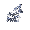
- Structure visualization
| Structure viewer | Molecule: MolmilJmol/JSmol |
|---|
- Downloads & links
-Download
| PDBx/mmCIF format | 1lg7.cif.gz | 48.8 KB | Display | PDBx/mmCIF format |
|---|---|---|---|---|
| PDB format | pdb1lg7.ent.gz | 34.8 KB | Display | PDB format |
| PDBx/mmJSON format | 1lg7.json.gz | Tree view | PDBx/mmJSON format | |
| Others |  Other downloads Other downloads |
-Validation report
| Arichive directory | https://data.pdbj.org/pub/pdb/validation_reports/lg/1lg7ftp://data.pdbj.org/pub/pdb/validation_reports/lg/1lg7 | HTTPS FTP |
|---|
-Related structure data
| Similar structure data |
|---|


-Links
 PDBj
PDBj- Assembly
| Deposited unit | 
| ||||||||
|---|---|---|---|---|---|---|---|---|---|
| 1 |
| ||||||||
| 2 | 
| ||||||||
| Unit cell |
|
-Components
| #1: Protein | Mass: 21011.041 Da / Num. of mol.: 1 / Source method: isolated from a natural source / Source: (natural) Vesicular stomatitis virus / Genus: Vesiculovirus / Strain: Indiana - Orsay / References: UniProt: Q8B0H2 |
|---|---|
| #2: Water | ChemComp-HOH /  Mass: 18.015 Da / Num. of mol.: 115 / Source method: isolated from a natural source / Formula: H2O Mass: 18.015 Da / Num. of mol.: 115 / Source method: isolated from a natural source / Formula: H2O |
| Has protein modification | Y |
-Experimental details
-Experiment
| Experiment | Method: X-RAY DIFFRACTION / Number of used crystals: 1 |
|---|
- Sample preparation
| Crystal | Density Matthews: 2 Å3/Da / Density % sol: 38.55 % | ||||||||||||||||||||||||||||||||||||||||||||||||||||||||
|---|---|---|---|---|---|---|---|---|---|---|---|---|---|---|---|---|---|---|---|---|---|---|---|---|---|---|---|---|---|---|---|---|---|---|---|---|---|---|---|---|---|---|---|---|---|---|---|---|---|---|---|---|---|---|---|---|---|
| Crystal grow | Temperature: 291 K / Method: vapor diffusion, hanging drop / pH: 8.5 Details: PEG 2000 monometylether, sodium chloride, sodium acetate, Tris, pH 8.5, VAPOR DIFFUSION, HANGING DROP, temperature 291K | ||||||||||||||||||||||||||||||||||||||||||||||||||||||||
| Crystal grow | *PLUS Details: Gaudier, M., (2001) Virology, 288, 308. | ||||||||||||||||||||||||||||||||||||||||||||||||||||||||
| Components of the solutions | *PLUS
|
-Data collection
| Diffraction | Mean temperature: 200 K |
|---|---|
| Diffraction source | Source: SYNCHROTRON / Site: ESRF  / Beamline: BM30A / Wavelength: 1 Å / Beamline: BM30A / Wavelength: 1 Å |
| Detector | Type: MAR scanner 345 mm plate / Detector: IMAGE PLATE / Date: Sep 17, 1999 |
| Radiation | Monochromator: Si 111 CHANNEL / Protocol: SINGLE WAVELENGTH / Monochromatic (M) / Laue (L): M / Scattering type: x-ray |
| Radiation wavelength | Wavelength: 1 Å / Relative weight: 1 |
| Reflection | Resolution: 1.96→25 Å / Num. all: 210584 / Num. obs: 209741 / % possible obs: 99.6 % / Observed criterion σ(F): 0 / Observed criterion σ(I): 0 / Rsym value: 0.069 / Net I/σ(I): 30 |
| Reflection shell | Resolution: 1.96→2.01 Å / Mean I/σ(I) obs: 3.5 / Rsym value: 0.41 / % possible all: 95.3 |
| Reflection | *PLUS Lowest resolution: 25 Å / Num. obs: 12771 / Num. measured all: 209741 / Rmerge(I) obs: 0.069 |
| Reflection shell | *PLUS % possible obs: 95.3 % / Rmerge(I) obs: 0.41 |
- Processing
| Software |
| ||||||||||||||||||||||||||||||||||||||||||||||||||||||||||||
|---|---|---|---|---|---|---|---|---|---|---|---|---|---|---|---|---|---|---|---|---|---|---|---|---|---|---|---|---|---|---|---|---|---|---|---|---|---|---|---|---|---|---|---|---|---|---|---|---|---|---|---|---|---|---|---|---|---|---|---|---|---|
| Refinement | Method to determine structure: SIRAS / Resolution: 1.96→25 Å / Cross valid method: THROUGHOUT / σ(F): 0 / Stereochemistry target values: Engh & Huber
| ||||||||||||||||||||||||||||||||||||||||||||||||||||||||||||
| Solvent computation | Bsol: 44.7872 Å2 / ksol: 0.335821 e/Å3 | ||||||||||||||||||||||||||||||||||||||||||||||||||||||||||||
| Displacement parameters | Biso mean: 29.7033 Å2 | ||||||||||||||||||||||||||||||||||||||||||||||||||||||||||||
| Refinement step | Cycle: LAST / Resolution: 1.96→25 Å
| ||||||||||||||||||||||||||||||||||||||||||||||||||||||||||||
| Refine LS restraints |
| ||||||||||||||||||||||||||||||||||||||||||||||||||||||||||||
| LS refinement shell | Resolution: 1.96→2.03 Å / Total num. of bins used: 10
| ||||||||||||||||||||||||||||||||||||||||||||||||||||||||||||
| Refinement | *PLUS Lowest resolution: 25 Å / Num. reflection obs: 12032 / Rfactor all: 0.218 / Rfactor obs: 0.206 / Rfactor Rfree: 0.241 / Rfactor Rwork: 0.206 | ||||||||||||||||||||||||||||||||||||||||||||||||||||||||||||
| Solvent computation | *PLUS | ||||||||||||||||||||||||||||||||||||||||||||||||||||||||||||
| Displacement parameters | *PLUS | ||||||||||||||||||||||||||||||||||||||||||||||||||||||||||||
| Refine LS restraints | *PLUS
| ||||||||||||||||||||||||||||||||||||||||||||||||||||||||||||
| LS refinement shell | *PLUS Rfactor Rfree: 0.327 / Rfactor Rwork: 0.239 / Rfactor obs: 0.239 |
