 Movie
Movie Controller

[English] 日本語
 Yorodumi
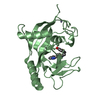
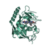
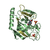
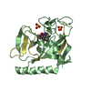
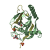
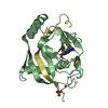
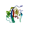
- PDB-1lfb: THE X-RAY STRUCTURE OF AN ATYPICAL HOMEODOMAIN PRESENT IN THE RAT... -
+ Open data

- Basic information
| Entry | Database: PDB / ID: 1lfb | ||||||
|---|---|---|---|---|---|---|---|
| Title | THE X-RAY STRUCTURE OF AN ATYPICAL HOMEODOMAIN PRESENT IN THE RAT LIVER TRANSCRIPTION FACTOR LFB1(SLASH)HNF1 AND IMPLICATIONS FOR DNA BINDING | ||||||
 Components Components | LIVER TRANSCRIPTION FACTOR (LFB1) | ||||||
 Keywords Keywords | TRANSCRIPTION REGULATION | ||||||
| Function / homology |  Function and homology information Function and homology informationparaxial mesoderm formation / apoptotic nuclear changes / regulation of NADP metabolic process / renal D-glucose absorption / D-glucose import across plasma membrane / cellular response to rapamycin / regulation of hormone secretion / activation of protein kinase activity / reproductive structure development / bile acid biosynthetic process ...paraxial mesoderm formation / apoptotic nuclear changes / regulation of NADP metabolic process / renal D-glucose absorption / D-glucose import across plasma membrane / cellular response to rapamycin / regulation of hormone secretion / activation of protein kinase activity / reproductive structure development / bile acid biosynthetic process / negative regulation of peptidyl-threonine phosphorylation / reverse cholesterol transport / cellular response to L-leucine / pancreas development / pronucleus / negative regulation of miRNA processing / regulation of Wnt signaling pathway / embryonic limb morphogenesis / heme biosynthetic process / insulin secretion / positive regulation of mitochondrial membrane potential / : / bile acid and bile salt transport / regulation of protein phosphorylation / positive regulation of ATP biosynthetic process / bone resorption / photoreceptor outer segment / blastocyst development / positive regulation of transcription initiation by RNA polymerase II / fatty acid transport / response to glucose / cholesterol metabolic process / placenta development / transcription coregulator binding / liver development / cellular response to glucose stimulus / transcription coactivator binding / fatty acid biosynthetic process / positive regulation of insulin secretion / intracellular protein localization / glucose homeostasis / response to oxidative stress / double-stranded DNA binding / DNA-binding transcription activator activity, RNA polymerase II-specific / transcription regulator complex / transcription by RNA polymerase II / sequence-specific DNA binding / DNA-binding transcription factor activity, RNA polymerase II-specific / positive regulation of phosphatidylinositol 3-kinase/protein kinase B signal transduction / protein dimerization activity / transcription cis-regulatory region binding / RNA polymerase II cis-regulatory region sequence-specific DNA binding / chromatin remodeling / DNA-binding transcription factor activity / protein heterodimerization activity / chromatin binding / positive regulation of gene expression / regulation of transcription by RNA polymerase II / regulation of DNA-templated transcription / negative regulation of apoptotic process / positive regulation of DNA-templated transcription / chromatin / negative regulation of transcription by RNA polymerase II / protein homodimerization activity / positive regulation of transcription by RNA polymerase II / protein-containing complex / DNA binding / nucleoplasm / identical protein binding / nucleus / cytoplasm Similarity search - Function | ||||||
| Biological species |  | ||||||
| Method |  X-RAY DIFFRACTION / Resolution: 2.8 Å X-RAY DIFFRACTION / Resolution: 2.8 Å | ||||||
 Authors Authors | Ceska, T.A. / Lamers, M. / Monaci, P. / Nicosia, A. / Cortese, R. / Suck, D. | ||||||
 Citation Citation | Journal: EMBO J. / Year: 1993 Title: The X-ray structure of an atypical homeodomain present in the rat liver transcription factor LFB1/HNF1 and implications for DNA binding. Authors: Ceska, T.A. / Lamers, M. / Monaci, P. / Nicosia, A. / Cortese, R. / Suck, D. | ||||||
| History |
|
- Structure visualization
| Structure viewer | Molecule: MolmilJmol/JSmol |
|---|
- Downloads & links
-Download
| PDBx/mmCIF format | 1lfb.cif.gz | 26.9 KB | Display | PDBx/mmCIF format |
|---|---|---|---|---|
| PDB format | pdb1lfb.ent.gz | 17.3 KB | Display | PDB format |
| PDBx/mmJSON format | 1lfb.json.gz | Tree view | PDBx/mmJSON format | |
| Others |  Other downloads Other downloads |
-Validation report
| Arichive directory | https://data.pdbj.org/pub/pdb/validation_reports/lf/1lfbftp://data.pdbj.org/pub/pdb/validation_reports/lf/1lfb | HTTPS FTP |
|---|
-Related structure data
| Similar structure data |
|---|


-Links
 PDBj
PDBj


- Assembly
| Deposited unit | 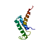
| ||||||||
|---|---|---|---|---|---|---|---|---|---|
| 1 | 
| ||||||||
| Unit cell |
|
-Components
| #1: Protein | Mass: 11677.199 Da / Num. of mol.: 1 Source method: isolated from a genetically manipulated source Source: (gene. exp.) |
|---|
-Experimental details
-Experiment
| Experiment | Method: X-RAY DIFFRACTION |
|---|
- Sample preparation
| Crystal | Density Matthews: 3.46 Å3/Da / Density % sol: 64.4 % | |||||||||||||||
|---|---|---|---|---|---|---|---|---|---|---|---|---|---|---|---|---|
| Crystal | *PLUS Density % sol: 65 % | |||||||||||||||
| Crystal grow | *PLUS pH: 5 / Method: vapor diffusion, hanging drop | |||||||||||||||
| Components of the solutions | *PLUS
|
-Data collection
| Radiation | Scattering type: x-ray |
|---|---|
| Radiation wavelength | Relative weight: 1 |
| Reflection | *PLUS Highest resolution: 2.8 Å / Num. all: 43872 / Num. obs: 4344 |
- Processing
| Software |
| ||||||||||||||||||||||||||||||||||||||||||||||||||||||||||||
|---|---|---|---|---|---|---|---|---|---|---|---|---|---|---|---|---|---|---|---|---|---|---|---|---|---|---|---|---|---|---|---|---|---|---|---|---|---|---|---|---|---|---|---|---|---|---|---|---|---|---|---|---|---|---|---|---|---|---|---|---|---|
| Refinement | Resolution: 2.8→6 Å
| ||||||||||||||||||||||||||||||||||||||||||||||||||||||||||||
| Refinement step | Cycle: LAST / Resolution: 2.8→6 Å
| ||||||||||||||||||||||||||||||||||||||||||||||||||||||||||||
| Refine LS restraints |
| ||||||||||||||||||||||||||||||||||||||||||||||||||||||||||||
| Refinement | *PLUS Highest resolution: 2.8 Å / Lowest resolution: 6 Å / Num. reflection obs: 2487 / Rfactor obs: 0.212 / Rfactor Rfree: 0.364 | ||||||||||||||||||||||||||||||||||||||||||||||||||||||||||||
| Solvent computation | *PLUS | ||||||||||||||||||||||||||||||||||||||||||||||||||||||||||||
| Displacement parameters | *PLUS | ||||||||||||||||||||||||||||||||||||||||||||||||||||||||||||
| Refine LS restraints | *PLUS Type: x_angle_d / Dev ideal: 3.36 |
