 ムービー
ムービー コントローラー


+ データを開く

- 基本情報
基本情報
| 登録情報 | データベース: PDB / ID: 1le1 | |||||||||
|---|---|---|---|---|---|---|---|---|---|---|
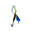
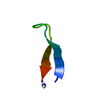
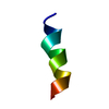
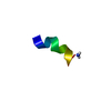
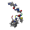
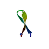
| タイトル | NMR Structure of Tryptophan Zipper 2: A stable, Monomeric Beta-Hairpin with a Type I' Turn | |||||||||
 要素 要素 | Tryptophan Zipper 2 | |||||||||
 キーワード キーワード | DE NOVO PROTEIN / beta-hairpin / type I' turn | |||||||||
| 手法 | 溶液NMR / Hybrid distance geometry, simulated annealing using the program DGII, molecular dynamics using the program AMBER, in conjunction not only with distance, dihedral angle restraints, but also 1H chemical shift-based restraints. | |||||||||
 データ登録者 データ登録者 | Cochran, A.G. / Skelton, N.J. / Starovasnik, M.A. | |||||||||
 引用 引用 | ジャーナル: Proc.Natl.Acad.Sci.USA / 年: 2001 タイトル: Tryptophan zippers: stable, monomeric beta -hairpins. 著者: Cochran, A.G. / Skelton, N.J. / Starovasnik, M.A. #1: ジャーナル: To be Publishedタイトル: Chemical-shift-based methods for structure validation and refinement: Application to tryptophan zipper beta-hairpin peptides 著者: Skelton, N.J. / Cochran, A.G. / Starovasnik, M.A. | |||||||||
| 履歴 |
|
- 構造の表示
構造の表示
| 構造ビューア | 分子:  MolmilJmol/JSmol MolmilJmol/JSmol |
|---|
- ダウンロードとリンク
-ダウンロード
| PDBx/mmCIF形式 | 1le1.cif.gz | 77.1 KB | 表示 | PDBx/mmCIF形式 |
|---|---|---|---|---|
| PDB形式 | pdb1le1.ent.gz | 60 KB | 表示 | PDB形式 |
| PDBx/mmJSON形式 | 1le1.json.gz | ツリー表示 | PDBx/mmJSON形式 | |
| その他 |  その他のダウンロード その他のダウンロード |
-検証レポート
| 文書・要旨 | 1le1_validation.pdf.gz | 332.9 KB | 表示 | wwPDB検証レポート |
|---|---|---|---|---|
| 文書・詳細版 | 1le1_full_validation.pdf.gz | 413.6 KB | 表示 | |
| XML形式データ | 1le1_validation.xml.gz | 5.2 KB | 表示 | |
| CIF形式データ | 1le1_validation.cif.gz | 8.2 KB | 表示 | |
| アーカイブディレクトリ | https://data.pdbj.org/pub/pdb/validation_reports/le/1le1ftp://data.pdbj.org/pub/pdb/validation_reports/le/1le1 | HTTPS FTP |
-関連構造データ


-リンク
 PDBj
PDBj
- 集合体
集合体
| 登録構造単位 | 
| |||||||||
|---|---|---|---|---|---|---|---|---|---|---|
| 1 |
| |||||||||
| NMR アンサンブル |
|
-要素
| #1: タンパク質・ペプチド | 分子量: 1608.776 Da / 分子数: 1 / 由来タイプ: 合成 詳細: This sequence was designed based on experimental data obtained from a model system. The peptide was chemically synthesized and does not appear to occur in nature. |
|---|
-実験情報
-実験
| 実験 | 手法: 溶液NMR | ||||||||||||||||||||||||
|---|---|---|---|---|---|---|---|---|---|---|---|---|---|---|---|---|---|---|---|---|---|---|---|---|---|
| NMR実験 |
| ||||||||||||||||||||||||
| NMR実験の詳細 | Text: The structure of trpzip2 was originally determined using standard 2D homonuclear techniques (Cochran et al., 2001, Proc. Natl. Acad. Sci. USA, 98, 5578-5583). Distance and dihedral angle ...Text: The structure of trpzip2 was originally determined using standard 2D homonuclear techniques (Cochran et al., 2001, Proc. Natl. Acad. Sci. USA, 98, 5578-5583). Distance and dihedral angle restraints were derived from analysis of NOESY, ROESY, DQF-COSY, and COSY-35 data. Chi-1 rotamers and stereospecific assignments for beta-methylene groups were based on combined analysis of 3JHaHb and local ROEs, suggesting that all four Trp residues adopt a chi1 of -60 deg. Quantitative analysis of the 1H chemical shifts, however, revealed that the frequencies of the Hb and He3 resonances of Trp4 and Trp11 were inconsistent with a -60 deg chi1 value, and indicated that the side chains for these tryptophans actually reside primarily in the 180 deg chi1 rotamer (Skelton et al., manuscript in preparation). The current coordinates result from refinement with the Sander module of AMBER (v6.0), and included not only NOE-derived distance restraints and dihedral angle restraints, but also 1H chemical shift-based restraints. Thus, the resulting updated coordinates differ from the previous ones in the side chain orientations of Trp4 and Trp11. However, the rest of the structure is similar with a backbone rmsd between the average coordinates of the two ensembles of 0.69 angstrom(res.2-11). The two strands are highly twisted, as described previously, however there is a slight difference in the relative position of the turn with respect to the strands. The key difference, however, is that each pair of cross-strand tryptophan rings now shows edge-to-face packing against one another. |
- 試料調製
試料調製
| 詳細 | 内容: 3mM trpzip2 / 溶媒系: 92% H2O, 8% D2O, pH 5.5, 0.1mM DSS |
|---|---|
| 試料状態 | イオン強度: 0 / pH: 5.5 / 圧: ambient / 温度: 288 K |
| 結晶化 | *PLUS 手法: other / 詳細: NMR |
-NMR測定
| 放射 | プロトコル: SINGLE WAVELENGTH / 単色(M)・ラウエ(L): M |
|---|---|
| 放射波長 | 相対比: 1 |
| NMRスペクトロメーター | タイプ: Bruker DRX / 製造業者: Bruker / モデル: DRX / 磁場強度: 500 MHz |
- 解析
解析
| NMR software |
| ||||||||||||||||||||
|---|---|---|---|---|---|---|---|---|---|---|---|---|---|---|---|---|---|---|---|---|---|
| 精密化 | 手法: Hybrid distance geometry, simulated annealing using the program DGII, molecular dynamics using the program AMBER, in conjunction not only with distance, dihedral angle restraints, but also 1H ...手法: Hybrid distance geometry, simulated annealing using the program DGII, molecular dynamics using the program AMBER, in conjunction not only with distance, dihedral angle restraints, but also 1H chemical shift-based restraints. ソフトェア番号: 1 詳細: Structures are based on a total of 80 (including 26 intra-residue, 15 sequential, 6 medium-range, and 33 long-range) NOE-derived distance restraints, 15 dihedral angle restraints, and 1H ...詳細: Structures are based on a total of 80 (including 26 intra-residue, 15 sequential, 6 medium-range, and 33 long-range) NOE-derived distance restraints, 15 dihedral angle restraints, and 1H chemical shift restraints for 39 carbon-bound 1H resonances; (chemical shift restraints were not imposed for the terminal residues, or for side chains exhibiting evidence of rotational averaging). The ensemble agrees well with the experimental restraints with no distance or dihedral angle violations greater than 0.10 angstrom or 5 deg, respectively, and an average rmsd of only 0.087 ppm between observed and calculated chemical shifts. | ||||||||||||||||||||
| 代表構造 | 選択基準: closest to the average | ||||||||||||||||||||
| NMRアンサンブル | コンフォーマー選択の基準: structures with the least restraint violations 計算したコンフォーマーの数: 76 / 登録したコンフォーマーの数: 20 |
 Amber
Amber