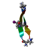
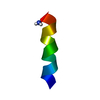
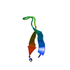
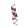
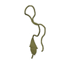
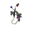
Mass: 1867.177 Da / Num. of mol.: 1 / Source method: obtained synthetically Details: The peptide was chemically synthesized. It is a novel sequence derived from phage-display selection.
Has protein modification
Y
-
Experimental details
-
Experiment
Experiment
Method: SOLUTION NMR
NMR experiment
Conditions-ID
Experiment-ID
Solution-ID
Type
1
1
1
2D-ROESY
1
2
1
2D-NOESY
1
3
1
DQF-COSY
2
4
2
COSY-35
2
5
2
2D-NOESY
2
6
2
2D-ROESY
NMR details
Text: This structure was determined using standard 2D homonuclear techniques. The sample contains a mixture of cis and trans isomers about the Gly7-Pro8 peptide bond. Both sets of resonances were ...Text: This structure was determined using standard 2D homonuclear techniques. The sample contains a mixture of cis and trans isomers about the Gly7-Pro8 peptide bond. Both sets of resonances were assigned. The trans isoform is not well ordered in solution. The cis isoform is structured, especially within the disulfide cycle. Structures were calculated on the basis of restraints generated only from the cis isoform.
-
Sample preparation
Details
Solution-ID
Contents
Solvent system
1
3mMpeptide
90% H2O, 10% D2O, pH5.1
2
3mMpeptide
100 % D2OpH5.1
Sample conditions
Conditions-ID
Ionic strength
pH
Pressure (kPa)
Temperature (K)
1
0
5.1
ambient
303K
2
0
5.1
ambient
303K
Crystal grow
*PLUS
Method: other / Details: NMR
-
NMR measurement
Radiation
Protocol: SINGLE WAVELENGTH / Monochromatic (M) / Laue (L): M
Radiation wavelength
Relative weight: 1
NMR spectrometer
Type: Bruker AVANCE / Manufacturer: Bruker / Model: AVANCE / Field strength: 500 MHz
-
Processing
NMR software
Name
Version
Developer
Classification
DGII
98
TimothyHavel
structuresolution
Discover
3.1
Accelrys
refinement
Refinement
Method: distance geometry, restrained molecular dynamics / Software ordinal: 1 Details: The structures are based on 52 NOE distance restraints, 11 phi and 4 chi-1 dihedral angle restraints. No hydrogen bond restraints were employed. The mean backbone atom RMSD to the mean ...Details: The structures are based on 52 NOE distance restraints, 11 phi and 4 chi-1 dihedral angle restraints. No hydrogen bond restraints were employed. The mean backbone atom RMSD to the mean structure within the disulfide cycle is 0.43 +/- 0.12 Angstoms.
NMR representative
Selection criteria: closest to the average
NMR ensemble
Conformer selection criteria: structures with the least restraint violations Conformers calculated total number: 80 / Conformers submitted total number: 20
+
About Yorodumi
-
News
-
Feb 9, 2022. New format data for meta-information of EMDB entries
New format data for meta-information of EMDB entries
Version 3 of the EMDB header file is now the official format.
The previous official version 1.9 will be removed from the archive.
In the structure databanks used in Yorodumi, some data are registered as the other names, "COVID-19 virus" and "2019-nCoV". Here are the details of the virus and the list of structure data.
Jan 31, 2019. EMDB accession codes are about to change! (news from PDBe EMDB page)
EMDB accession codes are about to change! (news from PDBe EMDB page)
The allocation of 4 digits for EMDB accession codes will soon come to an end. Whilst these codes will remain in use, new EMDB accession codes will include an additional digit and will expand incrementally as the available range of codes is exhausted. The current 4-digit format prefixed with “EMD-” (i.e. EMD-XXXX) will advance to a 5-digit format (i.e. EMD-XXXXX), and so on. It is currently estimated that the 4-digit codes will be depleted around Spring 2019, at which point the 5-digit format will come into force.
The EM Navigator/Yorodumi systems omit the EMD- prefix.
Related info.:Q: What is EMD? / ID/Accession-code notation in Yorodumi/EM Navigator
Yorodumi is a browser for structure data from EMDB, PDB, SASBDB, etc.
This page is also the successor to EM Navigator detail page, and also detail information page/front-end page for Omokage search.
The word "yorodu" (or yorozu) is an old Japanese word meaning "ten thousand". "mi" (miru) is to see.
Related info.:EMDB / PDB / SASBDB / Comparison of 3 databanks / Yorodumi Search / Aug 31, 2016. New EM Navigator & Yorodumi / Yorodumi Papers / Jmol/JSmol / Function and homology information / Changes in new EM Navigator and Yorodumi
 Movie
Movie Controller


 Open data
 Basic information
 Components
 Keywords
 Authors
 Citation
 Structure visualization
 Molmil
 Downloads & links
 Other downloads


 PDBj
PDBj
 Assembly
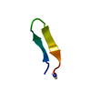
 Sample preparation
 Processing