 Movie
Movie Controller

[English] 日本語
 Yorodumi
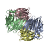
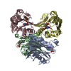
- PDB-1kuu: CRYSTAL STRUCTURE OF METHANOBACTERIUM THERMOAUTOTROPHICUM CONSERV... -
+ Open data

- Basic information
| Entry | Database: PDB / ID: 1kuu | ||||||
|---|---|---|---|---|---|---|---|
| Title | CRYSTAL STRUCTURE OF METHANOBACTERIUM THERMOAUTOTROPHICUM CONSERVED PROTEIN MTH1020 REVEALS AN NTN-HYDROLASE FOLD | ||||||
 Components Components | conserved protein | ||||||
 Keywords Keywords | STRUCTURAL GENOMICS / UNKNOWN FUNCTION / conserved protein / NTN-hydrolase fold | ||||||
| Function / homology |  Function and homology information Function and homology informationIMP cyclohydrolase / IMP cyclohydrolase activity / 'de novo' IMP biosynthetic process Similarity search - Function | ||||||
| Biological species |   Methanothermobacter (archaea) Methanothermobacter (archaea) | ||||||
| Method |  X-RAY DIFFRACTION / SYNCHROTRON / MAD / Resolution: 2.2 Å X-RAY DIFFRACTION / SYNCHROTRON / MAD / Resolution: 2.2 Å | ||||||
 Authors Authors | Saridakis, V. / Christendat, D. / Thygesen, A. / Arrowsmith, C.H. / Edwards, A.M. / Pai, E.F. | ||||||
 Citation Citation | Journal: PROTEINS: STRUCT.,FUNCT.,GENET. / Year: 2002 Title: CRYSTAL STRUCTURE OF METHANOBACTERIUM THERMOAUTOTROPHICUM CONSERVED PROTEIN MTH1020 REVEALS AN NTN-HYDROLASE FOLD Authors: Saridakis, V. / Christendat, D. / Thygesen, A. / Arrowsmith, C.H. / Edwards, A.M. / Pai, E.F. | ||||||
| History |
|
- Structure visualization
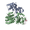
| Structure viewer | Molecule: MolmilJmol/JSmol |
|---|
- Downloads & links
-Download
| PDBx/mmCIF format | 1kuu.cif.gz | 49.9 KB | Display | PDBx/mmCIF format |
|---|---|---|---|---|
| PDB format | pdb1kuu.ent.gz | 36.2 KB | Display | PDB format |
| PDBx/mmJSON format | 1kuu.json.gz | Tree view | PDBx/mmJSON format | |
| Others |  Other downloads Other downloads |
-Validation report
| Arichive directory | https://data.pdbj.org/pub/pdb/validation_reports/ku/1kuuftp://data.pdbj.org/pub/pdb/validation_reports/ku/1kuu | HTTPS FTP |
|---|
-Related structure data
| Similar structure data |
|---|


-Links
 PDBj
PDBj- Assembly
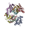
| Deposited unit | 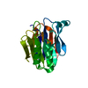
| ||||||||
|---|---|---|---|---|---|---|---|---|---|
| 1 | 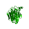
| ||||||||
| Unit cell |
|
-Components
| #1: Protein | Mass: 21859.414 Da / Num. of mol.: 1 Source method: isolated from a genetically manipulated source Source: (gene. exp.) Methanothermobacter (archaea) / Genus: Methanothermobacter / Gene: MTH1020 / Plasmid: pET15B / Species (production host): Escherichia coli / Production host:  |
|---|---|
| #2: Water | ChemComp-HOH /  Mass: 18.015 Da / Num. of mol.: 32 / Source method: isolated from a natural source / Formula: H2O Mass: 18.015 Da / Num. of mol.: 32 / Source method: isolated from a natural source / Formula: H2O |
-Experimental details
-Experiment
| Experiment | Method: X-RAY DIFFRACTION / Number of used crystals: 1 |
|---|
- Sample preparation
| Crystal | Density Matthews: 2.85 Å3/Da / Density % sol: 56.84 % | ||||||||||||||||||||||||
|---|---|---|---|---|---|---|---|---|---|---|---|---|---|---|---|---|---|---|---|---|---|---|---|---|---|
| Crystal grow | Temperature: 298 K / Method: vapor diffusion, hanging drop / pH: 7.5 Details: 14 % MPD, 0.2 M Mg Acetate, 100 mM HEPES, pH 7.5, VAPOR DIFFUSION, HANGING DROP, temperature 298K | ||||||||||||||||||||||||
| Crystal | *PLUS Density % sol: 57 % | ||||||||||||||||||||||||
| Crystal grow | *PLUS Temperature: 20 ℃ / Details: Christendat, D., (2000) J.Biol.Chem., 275, 24608. | ||||||||||||||||||||||||
| Components of the solutions | *PLUS
|
-Data collection
| Diffraction | Mean temperature: 100 K |
|---|---|
| Diffraction source | Source: SYNCHROTRON / Site: NSLS  / Beamline: X8C / Wavelength: 1 Å / Beamline: X8C / Wavelength: 1 Å |
| Detector | Type: ADSC QUANTUM 4 / Detector: CCD / Date: Aug 10, 2000 |
| Radiation | Protocol: SINGLE WAVELENGTH / Monochromatic (M) / Laue (L): M / Scattering type: x-ray |
| Radiation wavelength | Wavelength: 1 Å / Relative weight: 1 |
| Reflection | Resolution: 2.2→30 Å / Num. all: 13025 / Num. obs: 13025 / % possible obs: 99 % / Observed criterion σ(F): 0 / Redundancy: 9 % / Biso Wilson estimate: 29.9 Å2 / Rmerge(I) obs: 0.049 / Net I/σ(I): 35.6 |
| Reflection shell | Resolution: 2.2→2.34 Å / Rsym value: 0.374 / % possible all: 96.5 |
| Reflection | *PLUS Num. measured all: 113676 |
| Reflection shell | *PLUS Highest resolution: 2.25 Å / Lowest resolution: 2.33 Å / % possible obs: 96.5 % / Rmerge(I) obs: 0.374 / Mean I/σ(I) obs: 4.3 |
- Processing
| Software |
| ||||||||||||||||||||||||||||||||||||
|---|---|---|---|---|---|---|---|---|---|---|---|---|---|---|---|---|---|---|---|---|---|---|---|---|---|---|---|---|---|---|---|---|---|---|---|---|---|
| Refinement | Method to determine structure: MAD / Resolution: 2.2→14.5 Å / Rfactor Rfree error: 0.01 / Data cutoff high absF: 245271.05 / Data cutoff low absF: 0 / Isotropic thermal model: RESTRAINED / Cross valid method: THROUGHOUT / σ(F): 0
| ||||||||||||||||||||||||||||||||||||
| Solvent computation | Solvent model: FLAT MODEL / Bsol: 38.1196 Å2 / ksol: 0.349789 e/Å3 | ||||||||||||||||||||||||||||||||||||
| Displacement parameters | Biso mean: 46.8 Å2
| ||||||||||||||||||||||||||||||||||||
| Refine analyze |
| ||||||||||||||||||||||||||||||||||||
| Refinement step | Cycle: LAST / Resolution: 2.2→14.5 Å
| ||||||||||||||||||||||||||||||||||||
| Refine LS restraints |
| ||||||||||||||||||||||||||||||||||||
| LS refinement shell | Resolution: 2.2→2.34 Å / Rfactor Rfree error: 0.03 / Total num. of bins used: 6
| ||||||||||||||||||||||||||||||||||||
| Xplor file |
| ||||||||||||||||||||||||||||||||||||
| Refine LS restraints | *PLUS
| ||||||||||||||||||||||||||||||||||||
| LS refinement shell | *PLUS Rfactor obs: 0.27 |
