 Movie
Movie Controller

[English] 日本語
 Yorodumi
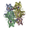
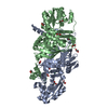
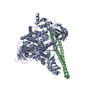
- PDB-1kp3: Crystal Structure of E. coli Argininosuccinate Synthetase in Comp... -
+ Open data

- Basic information
| Entry | Database: PDB / ID: 1kp3 | |||||||||
|---|---|---|---|---|---|---|---|---|---|---|
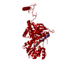
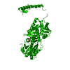
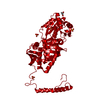
| Title | Crystal Structure of E. coli Argininosuccinate Synthetase in Complex with ATP and Citrulline | |||||||||
 Components Components | argininosuccinate synthetase | |||||||||
 Keywords Keywords | LIGASE / N-Type ATP pyrophosphatase | |||||||||
| Function / homology |  Function and homology information Function and homology informationargininosuccinate synthase / : / argininosuccinate synthase activity / L-arginine biosynthetic process / urea cycle / protein homodimerization activity / ATP binding / identical protein binding / cytoplasm / cytosol Similarity search - Function | |||||||||
| Biological species |  | |||||||||
| Method |  X-RAY DIFFRACTION / MOLECULAR REPLACEMENT / Resolution: 2 Å X-RAY DIFFRACTION / MOLECULAR REPLACEMENT / Resolution: 2 Å | |||||||||
 Authors Authors | Lemke, C.T. / Howell, P.L. | |||||||||
 Citation Citation | Journal: J.Biol.Chem. / Year: 2002 Title: Substrate Induced Conformational Changes in Argininosuccinate Synthetase Authors: Lemke, C.T. / Howell, P.L. #1: Journal: Structure / Year: 2001Title: The 1.6 A Crystal Structure of E. coli Argininosuccinate Synthetase Suggests a Conformational Change during Catalysis Authors: Lemke, C.T. / Howell, P.L. | |||||||||
| History |
|
- Structure visualization
| Structure viewer | Molecule: MolmilJmol/JSmol |
|---|
- Downloads & links
-Download
| PDBx/mmCIF format | 1kp3.cif.gz | 106.4 KB | Display | PDBx/mmCIF format |
|---|---|---|---|---|
| PDB format | pdb1kp3.ent.gz | 79.8 KB | Display | PDB format |
| PDBx/mmJSON format | 1kp3.json.gz | Tree view | PDBx/mmJSON format | |
| Others |  Other downloads Other downloads |
-Validation report
| Arichive directory | https://data.pdbj.org/pub/pdb/validation_reports/kp/1kp3ftp://data.pdbj.org/pub/pdb/validation_reports/kp/1kp3 | HTTPS FTP |
|---|
-Related structure data
| Related structure data |  1kp2C  1k92S S: Starting model for refinement C: citing same article ( |
|---|---|
| Similar structure data |


-Links
 PDBj
PDBj- Assembly
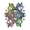
| Deposited unit | 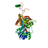
| ||||||||||||
|---|---|---|---|---|---|---|---|---|---|---|---|---|---|
| 1 | 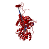
| ||||||||||||
| Unit cell |
| ||||||||||||
| Components on special symmetry positions |
| ||||||||||||
| Details | The biological assembly is a tetramer. |
-Components
-Protein , 1 types, 1 molecules A
| #1: Protein | Mass: 50967.262 Da / Num. of mol.: 1 Source method: isolated from a genetically manipulated source Source: (gene. exp.) |
|---|
-Non-polymers , 5 types, 192 molecules 


| #2: Chemical | ChemComp-PO4 /  Mass: 94.971 Da / Num. of mol.: 1 / Source method: obtained synthetically / Formula: PO4 Mass: 94.971 Da / Num. of mol.: 1 / Source method: obtained synthetically / Formula: PO4 | ||||
|---|---|---|---|---|---|
| #3: Chemical | ChemComp-ATP /  Mass: 507.181 Da / Num. of mol.: 1 / Source method: obtained synthetically / Formula: C10H16N5O13P3 / Comment: ATP, energy-carrying molecule*YM Mass: 507.181 Da / Num. of mol.: 1 / Source method: obtained synthetically / Formula: C10H16N5O13P3 / Comment: ATP, energy-carrying molecule*YM | ||||
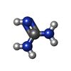
| #4: Chemical |  Type: L-peptide linking / Mass: 175.186 Da / Num. of mol.: 2 / Source method: obtained synthetically / Formula: C6H13N3O3 Type: L-peptide linking / Mass: 175.186 Da / Num. of mol.: 2 / Source method: obtained synthetically / Formula: C6H13N3O3#5: Chemical |  Mass: 59.070 Da / Num. of mol.: 2 / Source method: obtained synthetically / Formula: CH5N3 Mass: 59.070 Da / Num. of mol.: 2 / Source method: obtained synthetically / Formula: CH5N3#6: Water | ChemComp-HOH / | Mass: 18.015 Da / Num. of mol.: 186 / Source method: isolated from a natural source / Formula: H2O |
-Experimental details
-Experiment
| Experiment | Method: X-RAY DIFFRACTION / Number of used crystals: 1 |
|---|
- Sample preparation
| Crystal | Density Matthews: 2.52 Å3/Da / Density % sol: 40 % | ||||||||||||||||||||||||||||||||||||||||||||||||||||||||||||||||||||||
|---|---|---|---|---|---|---|---|---|---|---|---|---|---|---|---|---|---|---|---|---|---|---|---|---|---|---|---|---|---|---|---|---|---|---|---|---|---|---|---|---|---|---|---|---|---|---|---|---|---|---|---|---|---|---|---|---|---|---|---|---|---|---|---|---|---|---|---|---|---|---|---|
| Crystal grow | Temperature: 298 K / Method: vapor diffusion, hanging drop / pH: 6.5 Details: sodium/potassium phosphate, guanidine hydrochloride, MES, pH 6.5, VAPOR DIFFUSION, HANGING DROP, temperature 298K | ||||||||||||||||||||||||||||||||||||||||||||||||||||||||||||||||||||||
| Crystal grow | *PLUS Details: used microseeding | ||||||||||||||||||||||||||||||||||||||||||||||||||||||||||||||||||||||
| Components of the solutions | *PLUS
|
-Data collection
| Diffraction | Mean temperature: 100 K |
|---|---|
| Diffraction source | Source: ROTATING ANODE / Type: RIGAKU RU200 / Wavelength: 1.5418 Å |
| Detector | Type: MARRESEARCH / Detector: IMAGE PLATE |
| Radiation | Monochromator: GRAPHITE / Protocol: SINGLE WAVELENGTH / Monochromatic (M) / Laue (L): M / Scattering type: x-ray |
| Radiation wavelength | Wavelength: 1.5418 Å / Relative weight: 1 |
| Reflection | Resolution: 2→41 Å / Num. all: 35158 / Num. obs: 32696 / % possible obs: 93 % / Observed criterion σ(F): 0 / Observed criterion σ(I): 0 / Redundancy: 6.7 % / Biso Wilson estimate: 20.1 Å2 / Rmerge(I) obs: 0.082 / Net I/σ(I): 11.8 |
| Reflection shell | Resolution: 1.99→2.06 Å / Rmerge(I) obs: 0.396 / % possible all: 71.9 |
| Reflection | *PLUS Highest resolution: 1.99 Å / Lowest resolution: 41 Å / Num. obs: 34170 / % possible obs: 95.6 % / Num. measured all: 229900 / Rmerge(I) obs: 0.082 |
| Reflection shell | *PLUS % possible obs: 71.9 % / Rmerge(I) obs: 0.396 |
- Processing
| Software |
| ||||||||||||||||||||||||||||||||||||
|---|---|---|---|---|---|---|---|---|---|---|---|---|---|---|---|---|---|---|---|---|---|---|---|---|---|---|---|---|---|---|---|---|---|---|---|---|---|
| Refinement | Method to determine structure: MOLECULAR REPLACEMENT Starting model: PDB ENTRY 1K92 Resolution: 2→40.84 Å / Rfactor Rfree error: 0.004 / Data cutoff high absF: 452054.58 / Data cutoff low absF: 0 / Isotropic thermal model: RESTRAINED / Cross valid method: THROUGHOUT / σ(F): 0 / Stereochemistry target values: Engh & Huber
| ||||||||||||||||||||||||||||||||||||
| Solvent computation | Solvent model: FLAT MODEL / Bsol: 57.3294 Å2 / ksol: 0.407088 e/Å3 | ||||||||||||||||||||||||||||||||||||
| Displacement parameters | Biso mean: 35.4 Å2
| ||||||||||||||||||||||||||||||||||||
| Refine analyze |
| ||||||||||||||||||||||||||||||||||||
| Refinement step | Cycle: LAST / Resolution: 2→40.84 Å
| ||||||||||||||||||||||||||||||||||||
| Refine LS restraints |
| ||||||||||||||||||||||||||||||||||||
| LS refinement shell | Resolution: 2→2.13 Å / Rfactor Rfree error: 0.016 / Total num. of bins used: 6
| ||||||||||||||||||||||||||||||||||||
| Xplor file |
| ||||||||||||||||||||||||||||||||||||
| Refinement | *PLUS Highest resolution: 2 Å / Lowest resolution: 41 Å / % reflection Rfree: 10 % / Rfactor obs: 0.1916 / Rfactor Rfree: 0.2294 / Rfactor Rwork: 0.1916 | ||||||||||||||||||||||||||||||||||||
| Solvent computation | *PLUS | ||||||||||||||||||||||||||||||||||||
| Displacement parameters | *PLUS | ||||||||||||||||||||||||||||||||||||
| Refine LS restraints | *PLUS
| ||||||||||||||||||||||||||||||||||||
| LS refinement shell | *PLUS Rfactor Rfree: 0.332 / Rfactor Rwork: 0.283 |
