 Movie
Movie Controller

[English] 日本語
 Yorodumi
- PDB-1kkb: Complex of Escherichia coli Adenylosuccinate Synthetase with IMP ... -
+ Open data

- Basic information
| Entry | Database: PDB / ID: 1kkb | ||||||
|---|---|---|---|---|---|---|---|
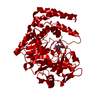
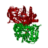
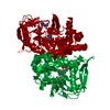
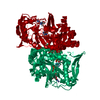
| Title | Complex of Escherichia coli Adenylosuccinate Synthetase with IMP and Hadacidin | ||||||
 Components Components | Adenylosuccinate Synthetase | ||||||
 Keywords Keywords | LIGASE / GTP-HYDROLYSING ENZYMES / PURINE NUCLEOTIDE / BIOSYNTHESIS / INDUCED FIT | ||||||
| Function / homology |  Function and homology information Function and homology informationadenylosuccinate synthase / adenylosuccinate synthase activity / adenosine biosynthetic process / IMP metabolic process / 'de novo' AMP biosynthetic process / nucleobase-containing small molecule interconversion / purine nucleotide biosynthetic process / guanosine tetraphosphate binding / DNA damage response / GTP binding ...adenylosuccinate synthase / adenylosuccinate synthase activity / adenosine biosynthetic process / IMP metabolic process / 'de novo' AMP biosynthetic process / nucleobase-containing small molecule interconversion / purine nucleotide biosynthetic process / guanosine tetraphosphate binding / DNA damage response / GTP binding / magnesium ion binding / membrane / cytoplasm / cytosol Similarity search - Function | ||||||
| Biological species |  | ||||||
| Method |  X-RAY DIFFRACTION / MOLECULAR REPLACEMENT / Resolution: 2.6 Å X-RAY DIFFRACTION / MOLECULAR REPLACEMENT / Resolution: 2.6 Å | ||||||
 Authors Authors | Hou, Z. / Wang, W. / Fromm, H.J. / Honzatko, R.B. | ||||||
 Citation Citation | Journal: J.Biol.Chem. / Year: 2002 Title: IMP Alone Organizes the Active Site of Adenylosuccinate Synthetase from Escherichia coli. Authors: Hou, Z. / Wang, W. / Fromm, H.J. / Honzatko, R.B. | ||||||
| History |
|
- Structure visualization
| Structure viewer | Molecule: MolmilJmol/JSmol |
|---|
- Downloads & links
-Download
| PDBx/mmCIF format | 1kkb.cif.gz | 102.4 KB | Display | PDBx/mmCIF format |
|---|---|---|---|---|
| PDB format | pdb1kkb.ent.gz | 77.4 KB | Display | PDB format |
| PDBx/mmJSON format | 1kkb.json.gz | Tree view | PDBx/mmJSON format | |
| Others |  Other downloads Other downloads |
-Validation report
| Arichive directory | https://data.pdbj.org/pub/pdb/validation_reports/kk/1kkbftp://data.pdbj.org/pub/pdb/validation_reports/kk/1kkb | HTTPS FTP |
|---|
-Related structure data


-Links
 PDBj
PDBj- Assembly
| Deposited unit | 
| ||||||||
|---|---|---|---|---|---|---|---|---|---|
| 1 | 
| ||||||||
| Unit cell |
| ||||||||
| Details | The asymmetry unit is one subunit. The functional synthetase is a dimer |
-Components
| #1: Protein | Mass: 47400.793 Da / Num. of mol.: 1 Source method: isolated from a genetically manipulated source Source: (gene. exp.) References: UniProt: P12283, UniProt: P0A7D4*PLUS, adenylosuccinate synthase |
|---|---|
| #2: Chemical | ChemComp-HDA /   Mass: 119.076 Da / Num. of mol.: 1 / Source method: obtained synthetically / Formula: C3H5NO4 / Comment: anticancer, medication*YM Mass: 119.076 Da / Num. of mol.: 1 / Source method: obtained synthetically / Formula: C3H5NO4 / Comment: anticancer, medication*YM |
| #3: Chemical | ChemComp-IMP /   Mass: 348.206 Da / Num. of mol.: 1 / Source method: obtained synthetically / Formula: C10H13N4O8P Mass: 348.206 Da / Num. of mol.: 1 / Source method: obtained synthetically / Formula: C10H13N4O8P |
| #4: Water | ChemComp-HOH /  Mass: 18.015 Da / Num. of mol.: 303 / Source method: isolated from a natural source / Formula: H2O Mass: 18.015 Da / Num. of mol.: 303 / Source method: isolated from a natural source / Formula: H2O |
-Experimental details
-Experiment
| Experiment | Method: X-RAY DIFFRACTION / Number of used crystals: 1 |
|---|
- Sample preparation
| Crystal | Density Matthews: 3.1 Å3/Da / Density % sol: 60.33 % | ||||||||||||||||||||||||||||||||||||||||||||||||||||||
|---|---|---|---|---|---|---|---|---|---|---|---|---|---|---|---|---|---|---|---|---|---|---|---|---|---|---|---|---|---|---|---|---|---|---|---|---|---|---|---|---|---|---|---|---|---|---|---|---|---|---|---|---|---|---|---|
| Crystal grow | Temperature: 293 K / Method: vapor diffusion, hanging drop / pH: 6.5 Details: PEG 8000, HEPES, IMP, Hadacidin, magnesium acetate, pH 6.5, VAPOR DIFFUSION, HANGING DROP, temperature 293K | ||||||||||||||||||||||||||||||||||||||||||||||||||||||
| Crystal grow | *PLUS pH: 6.8 | ||||||||||||||||||||||||||||||||||||||||||||||||||||||
| Components of the solutions | *PLUS
|
-Data collection
| Diffraction | Mean temperature: 120 K |
|---|---|
| Diffraction source | Source: ROTATING ANODE / Type: SIEMENS / Wavelength: 1.54178 Å |
| Detector | Type: SIEMENS / Detector: AREA DETECTOR / Date: Jan 1, 1998 / Details: Graphite monochromator |
| Radiation | Monochromator: Graphite / Protocol: SINGLE WAVELENGTH / Monochromatic (M) / Laue (L): M / Scattering type: x-ray |
| Radiation wavelength | Wavelength: 1.54178 Å / Relative weight: 1 |
| Reflection | Resolution: 2.6→20 Å / Num. all: 22708 / Num. obs: 22708 / % possible obs: 91 % / Observed criterion σ(F): 0 / Observed criterion σ(I): 0 / Redundancy: 5 % / Rsym value: 0.122 |
| Reflection shell | Resolution: 2.6→2.9 Å / % possible all: 90 |
| Reflection | *PLUS Lowest resolution: 20 Å / % possible obs: 91 % / Num. measured all: 121026 / Rmerge(I) obs: 0.12 |
| Reflection shell | *PLUS % possible obs: 90 % |
- Processing
| Software |
| |||||||||||||||||||||||||
|---|---|---|---|---|---|---|---|---|---|---|---|---|---|---|---|---|---|---|---|---|---|---|---|---|---|---|
| Refinement | Method to determine structure: MOLECULAR REPLACEMENT / Resolution: 2.6→5 Å / Cross valid method: THROUGHOUT / σ(F): 0
| |||||||||||||||||||||||||
| Refinement step | Cycle: LAST / Resolution: 2.6→5 Å
| |||||||||||||||||||||||||
| Refine LS restraints |
| |||||||||||||||||||||||||
| Refinement | *PLUS Lowest resolution: 5 Å / σ(F): 0 / % reflection Rfree: 10 % / Rfactor obs: 0.18 / Rfactor Rfree: 0.29 / Rfactor Rwork: 0.18 | |||||||||||||||||||||||||
| Solvent computation | *PLUS | |||||||||||||||||||||||||
| Displacement parameters | *PLUS | |||||||||||||||||||||||||
| Refine LS restraints | *PLUS
|
