 Movie
Movie Controller

[English] 日本語
 Yorodumi
- PDB-1kh1: Crystal Structure of Thermus thermophilus HB8 Argininosuccinate S... -
+ Open data

- Basic information
| Entry | Database: PDB / ID: 1kh1 | ||||||
|---|---|---|---|---|---|---|---|
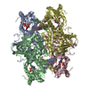
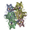
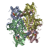
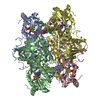
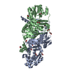
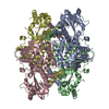
| Title | Crystal Structure of Thermus thermophilus HB8 Argininosuccinate Synthetase | ||||||
 Components Components | Argininosuccinate Synthetase | ||||||
 Keywords Keywords | LIGASE / RIKEN Structural Genomics/Proteomics Initiative / RSGI / Structural Genomics | ||||||
| Function / homology |  Function and homology information Function and homology informationargininosuccinate synthase / argininosuccinate metabolic process / argininosuccinate synthase activity / urea cycle / L-arginine biosynthetic process / ATP binding / cytoplasm Similarity search - Function | ||||||
| Biological species |   Thermus thermophilus (bacteria) Thermus thermophilus (bacteria) | ||||||
| Method |  X-RAY DIFFRACTION / SYNCHROTRON / MIR / Resolution: 2.3 Å X-RAY DIFFRACTION / SYNCHROTRON / MIR / Resolution: 2.3 Å | ||||||
 Authors Authors | Goto, M. / Nakajima, Y. / Hirotsu, K. / RIKEN Structural Genomics/Proteomics Initiative (RSGI) | ||||||
 Citation Citation | Journal: J.Biol.Chem. / Year: 2002 Title: Crystal structure of argininosuccinate synthetase from Thermus thermophilus HB8. Structural basis for the catalytic action. Authors: Goto, M. / Nakajima, Y. / Hirotsu, K. | ||||||
| History |
|
- Structure visualization
| Structure viewer | Molecule: MolmilJmol/JSmol |
|---|
- Downloads & links
-Download
| PDBx/mmCIF format | 1kh1.cif.gz | 309.2 KB | Display | PDBx/mmCIF format |
|---|---|---|---|---|
| PDB format | pdb1kh1.ent.gz | 253.5 KB | Display | PDB format |
| PDBx/mmJSON format | 1kh1.json.gz | Tree view | PDBx/mmJSON format | |
| Others |  Other downloads Other downloads |
-Validation report
| Summary document | 1kh1_validation.pdf.gz | 476.2 KB | Display | wwPDB validaton report |
|---|---|---|---|---|
| Full document | 1kh1_full_validation.pdf.gz | 515.8 KB | Display | |
| Data in XML | 1kh1_validation.xml.gz | 61.2 KB | Display | |
| Data in CIF | 1kh1_validation.cif.gz | 84 KB | Display | |
| Arichive directory | https://data.pdbj.org/pub/pdb/validation_reports/kh/1kh1ftp://data.pdbj.org/pub/pdb/validation_reports/kh/1kh1 | HTTPS FTP |
-Related structure data


-Links
 PDBj
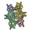
PDBj- Assembly
| Deposited unit | 
| ||||||||
|---|---|---|---|---|---|---|---|---|---|
| 1 |
| ||||||||
| Unit cell |
|
-Components
| #1: Protein | Mass: 44875.992 Da / Num. of mol.: 4 Source method: isolated from a genetically manipulated source Source: (gene. exp.) Thermus thermophilus (bacteria) / Gene: HB8 / Plasmid: pET11a / Production host: #2: Chemical | ChemComp-SO4 /   Mass: 96.063 Da / Num. of mol.: 4 / Source method: obtained synthetically / Formula: SO4 Mass: 96.063 Da / Num. of mol.: 4 / Source method: obtained synthetically / Formula: SO4#3: Water | ChemComp-HOH / |  Mass: 18.015 Da / Num. of mol.: 458 / Source method: isolated from a natural source / Formula: H2O Mass: 18.015 Da / Num. of mol.: 458 / Source method: isolated from a natural source / Formula: H2O |
|---|
-Experimental details
-Experiment
| Experiment | Method: X-RAY DIFFRACTION / Number of used crystals: 1 |
|---|
- Sample preparation
| Crystal | Density Matthews: 4.52 Å3/Da / Density % sol: 72.76 % | ||||||||||||||||||||||||||||||
|---|---|---|---|---|---|---|---|---|---|---|---|---|---|---|---|---|---|---|---|---|---|---|---|---|---|---|---|---|---|---|---|
| Crystal grow | Temperature: 293 K / Method: vapor diffusion, hanging drop / pH: 8.5 Details: tris, ammonium sulfate, glycerol, pH 8.5, VAPOR DIFFUSION, HANGING DROP, temperature 293K | ||||||||||||||||||||||||||||||
| Crystal grow | *PLUS | ||||||||||||||||||||||||||||||
| Components of the solutions | *PLUS
|
-Data collection
| Diffraction | Mean temperature: 100 K |
|---|---|
| Diffraction source | Source: SYNCHROTRON / Site: SPring-8  / Beamline: BL41XU / Wavelength: 1 Å / Beamline: BL41XU / Wavelength: 1 Å |
| Detector | Type: MARRESEARCH / Detector: CCD / Date: Nov 26, 2000 |
| Radiation | Protocol: SINGLE WAVELENGTH / Monochromatic (M) / Laue (L): M / Scattering type: x-ray |
| Radiation wavelength | Wavelength: 1 Å / Relative weight: 1 |
| Reflection | Resolution: 2.3→48.8 Å / Num. all: 124435 / Num. obs: 124435 / % possible obs: 89.2 % / Rmerge(I) obs: 0.065 |
| Reflection shell | Resolution: 2.3→2.37 Å / % possible all: 78.3 |
| Reflection | *PLUS Highest resolution: 2.3 Å / Num. measured all: 281918 / Rmerge(I) obs: 0.065 |
| Reflection shell | *PLUS % possible obs: 78.3 % / Rmerge(I) obs: 0.127 |
- Processing
| Software |
| |||||||||||||||||||||||||
|---|---|---|---|---|---|---|---|---|---|---|---|---|---|---|---|---|---|---|---|---|---|---|---|---|---|---|
| Refinement | Method to determine structure: MIR / Resolution: 2.3→10 Å / σ(F): 2
| |||||||||||||||||||||||||
| Refine analyze |
| |||||||||||||||||||||||||
| Refinement step | Cycle: LAST / Resolution: 2.3→10 Å
| |||||||||||||||||||||||||
| Refine LS restraints |
| |||||||||||||||||||||||||
| Refinement | *PLUS Highest resolution: 2.3 Å / Lowest resolution: 48.8 Å / Rfactor obs: 0.1994 / Rfactor Rfree: 0.2291 / Rfactor Rwork: 0.1994 | |||||||||||||||||||||||||
| Solvent computation | *PLUS | |||||||||||||||||||||||||
| Displacement parameters | *PLUS | |||||||||||||||||||||||||
| LS refinement shell | *PLUS Rfactor Rfree: 0.2907 / Rfactor Rwork: 0.2525 / Rfactor obs: 0.2525 |
