 Movie
Movie Controller

[English] 日本語
 Yorodumi
- PDB-1kcb: Crystal Structure of a NO-forming Nitrite Reductase Mutant: an An... -
+ Open data

- Basic information
| Entry | Database: PDB / ID: 1kcb | ||||||
|---|---|---|---|---|---|---|---|
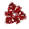
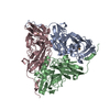
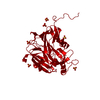
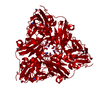
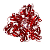
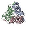
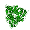
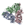
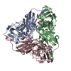
| Title | Crystal Structure of a NO-forming Nitrite Reductase Mutant: an Analog of a Transition State in Enzymatic Reaction | ||||||
 Components Components | Nitrite Reductase | ||||||
 Keywords Keywords | OXIDOREDUCTASE / copper-containing nitrite reductase / BETA BARREL | ||||||
| Function / homology |  Function and homology information Function and homology informationdenitrification pathway / nitrite reductase (NO-forming) / nitrite reductase (NO-forming) activity / nitrate assimilation / periplasmic space / copper ion binding Similarity search - Function | ||||||
| Biological species |  Achromobacter cycloclastes (bacteria) Achromobacter cycloclastes (bacteria) | ||||||
| Method |  X-RAY DIFFRACTION / SYNCHROTRON / FOURIER SYNTHESIS / Resolution: 1.65 Å X-RAY DIFFRACTION / SYNCHROTRON / FOURIER SYNTHESIS / Resolution: 1.65 Å | ||||||
 Authors Authors | Liu, S.Q. / Chang, T. / Liu, M.Y. / LeGall, J. / Chang, W.C. / Zhang, J.P. / Liang, D.C. / Chang, W.R. | ||||||
 Citation Citation | Journal: Biochem.Biophys.Res.Commun. / Year: 2003 Title: Crystal structure of a NO-forming nitrite reductase mutant: an analog of a transition state in enzymatic reaction Authors: Liu, S.Q. / Chang, T. / Liu, M.Y. / LeGall, J. / Chang, W.C. / Zhang, J.P. / Liang, D.C. / Chang, W.R. | ||||||
| History |
|
- Structure visualization
| Structure viewer | Molecule: MolmilJmol/JSmol |
|---|
- Downloads & links
-Download
| PDBx/mmCIF format | 1kcb.cif.gz | 81.5 KB | Display | PDBx/mmCIF format |
|---|---|---|---|---|
| PDB format | pdb1kcb.ent.gz | 59.5 KB | Display | PDB format |
| PDBx/mmJSON format | 1kcb.json.gz | Tree view | PDBx/mmJSON format | |
| Others |  Other downloads Other downloads |
-Validation report
| Summary document | 1kcb_validation.pdf.gz | 433.3 KB | Display | wwPDB validaton report |
|---|---|---|---|---|
| Full document | 1kcb_full_validation.pdf.gz | 437.5 KB | Display | |
| Data in XML | 1kcb_validation.xml.gz | 15.4 KB | Display | |
| Data in CIF | 1kcb_validation.cif.gz | 21.8 KB | Display | |
| Arichive directory | https://data.pdbj.org/pub/pdb/validation_reports/kc/1kcbftp://data.pdbj.org/pub/pdb/validation_reports/kc/1kcb | HTTPS FTP |
-Related structure data
| Related structure data |  1nieS S: Starting model for refinement |
|---|---|
| Similar structure data |

-Links
 PDBj
PDBj

- Assembly
| Deposited unit | 
| |||||||||
|---|---|---|---|---|---|---|---|---|---|---|
| 1 | 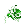
| |||||||||
| Unit cell |
| |||||||||
| Components on special symmetry positions |
| |||||||||
| Details | The biological assembly is a trimer generated from the monomer in the asymmetric unit by the operations: y, z, x and z, x, y |
-Components
| #1: Protein | Mass: 37075.766 Da / Num. of mol.: 1 / Mutation: I257E Source method: isolated from a genetically manipulated source Source: (gene. exp.) Achromobacter cycloclastes (bacteria) / Plasmid: pQE30 / Production host: | ||
|---|---|---|---|
| #2: Chemical |   Mass: 63.546 Da / Num. of mol.: 2 / Source method: obtained synthetically / Formula: Cu Mass: 63.546 Da / Num. of mol.: 2 / Source method: obtained synthetically / Formula: Cu#3: Water | ChemComp-HOH / |  Mass: 18.015 Da / Num. of mol.: 167 / Source method: isolated from a natural source / Formula: H2O Mass: 18.015 Da / Num. of mol.: 167 / Source method: isolated from a natural source / Formula: H2O |
-Experimental details
-Experiment
| Experiment | Method: X-RAY DIFFRACTION / Number of used crystals: 1 |
|---|
- Sample preparation
| Crystal | Density Matthews: 2.05 Å3/Da / Density % sol: 39.56 % | ||||||||||||||||||||||||||||||
|---|---|---|---|---|---|---|---|---|---|---|---|---|---|---|---|---|---|---|---|---|---|---|---|---|---|---|---|---|---|---|---|
| Crystal grow | Temperature: 293 K / Method: vapor diffusion, hanging drop / pH: 4 Details: ammonium sulfate, Potassium Phosphate, pH 4.0, VAPOR DIFFUSION, HANGING DROP, temperature 293K | ||||||||||||||||||||||||||||||
| Crystal grow | *PLUS Temperature: 15 ℃ / Method: vapor diffusion, hanging drop | ||||||||||||||||||||||||||||||
| Components of the solutions | *PLUS
|
-Data collection
| Diffraction | Mean temperature: 293 K |
|---|---|
| Diffraction source | Source: SYNCHROTRON / Site: Photon Factory  / Beamline: BL-6B / Wavelength: 1 Å / Beamline: BL-6B / Wavelength: 1 Å |
| Detector | Type: FUJI / Detector: IMAGE PLATE / Date: Nov 30, 1999 |
| Radiation | Protocol: SINGLE WAVELENGTH / Monochromatic (M) / Laue (L): M / Scattering type: x-ray |
| Radiation wavelength | Wavelength: 1 Å / Relative weight: 1 |
| Reflection | Resolution: 1.5→10 Å / Num. all: 37951 / Num. obs: 37951 / % possible obs: 100 % / Observed criterion σ(F): 0 / Observed criterion σ(I): 0 / Rmerge(I) obs: 0.052 / Net I/σ(I): 48.2 |
| Reflection shell | Resolution: 1.65→1.69 Å / Rmerge(I) obs: 0.25 / Mean I/σ(I) obs: 3.7 / % possible all: 100 |
| Reflection | *PLUS Highest resolution: 1.65 Å / Num. obs: 37954 / % possible obs: 99.9 % / Num. measured all: 393521 |
| Reflection shell | *PLUS Mean I/σ(I) obs: 3 |
- Processing
| Software |
| |||||||||||||||||||||||||
|---|---|---|---|---|---|---|---|---|---|---|---|---|---|---|---|---|---|---|---|---|---|---|---|---|---|---|
| Refinement | Method to determine structure: FOURIER SYNTHESIS Starting model: PDB ENTRY 1NIE Resolution: 1.65→9.97 Å / σ(F): 4 / σ(I): 2
| |||||||||||||||||||||||||
| Refinement step | Cycle: LAST / Resolution: 1.65→9.97 Å
| |||||||||||||||||||||||||
| Refine LS restraints |
| |||||||||||||||||||||||||
| Refinement | *PLUS Lowest resolution: 10 Å / Rfactor Rwork: 0.17 | |||||||||||||||||||||||||
| Solvent computation | *PLUS | |||||||||||||||||||||||||
| Displacement parameters | *PLUS |
