 Movie
Movie Controller

[English] 日本語
 Yorodumi
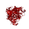
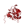
- PDB-1kbw: CRYSTAL STRUCTURE OF THE SOLUBLE DOMAIN OF ANIA FROM NEISSERIA GO... -
+ Open data

- Basic information
| Entry | Database: PDB / ID: 1kbw | ||||||
|---|---|---|---|---|---|---|---|
| Title | CRYSTAL STRUCTURE OF THE SOLUBLE DOMAIN OF ANIA FROM NEISSERIA GONORRHOEAE | ||||||
 Components Components | Major outer membrane protein PAN 1 | ||||||
 Keywords Keywords | OXIDOREDUCTASE / ANIA | ||||||
| Function / homology |  Function and homology information Function and homology informationnitrite reductase (NO-forming) / nitrite reductase (NO-forming) activity / cell outer membrane / copper ion binding Similarity search - Function | ||||||
| Biological species |  Neisseria gonorrhoeae (bacteria) Neisseria gonorrhoeae (bacteria) | ||||||
| Method |  X-RAY DIFFRACTION / SYNCHROTRON / MOLECULAR REPLACEMENT / Resolution: 2.4 Å X-RAY DIFFRACTION / SYNCHROTRON / MOLECULAR REPLACEMENT / Resolution: 2.4 Å | ||||||
 Authors Authors | Boulanger, M.J. / Murphy, M.E.P. | ||||||
 Citation Citation | Journal: J.Mol.Biol. / Year: 2002 Title: Crystal structure of the soluble domain of the major anaerobically induced outer membrane protein (AniA) from pathogenic Neisseria: a new class of copper-containing nitrite reductases. Authors: Boulanger, M.J. / Murphy, M.E. | ||||||
| History |
| ||||||
| Remark 999 | SEQUENCE ACCORDING TO THE AUTHOR THE SEQUENCE DIFFERENCES EXIST BETWEEN HIS SEQUENCE AND THE ...SEQUENCE ACCORDING TO THE AUTHOR THE SEQUENCE DIFFERENCES EXIST BETWEEN HIS SEQUENCE AND THE SWISSPROT ENTRY Q02219. AUTHOR'S SEQUENCE IS IDENTICAL TO THE SEQUENCE OBTAINGED FROM THE GONOCOCCAL GENOME SEQUENCING PROJECT (A49208) SUPPORTED BY USPHS/NIH GRANT #AI38399, AND B.A.ROE, L.SONG, S.P.LIN, X.YUAN, S.CLIFTON, T.DUCEY, L.LEWIS AND D.W.DYER AT THE UNIVERSITY OF OKLAHOMA - ACGT. |
- Structure visualization
| Structure viewer | Molecule: MolmilJmol/JSmol |
|---|
- Downloads & links
-Download
| PDBx/mmCIF format | 1kbw.cif.gz | 369.5 KB | Display | PDBx/mmCIF format |
|---|---|---|---|---|
| PDB format | pdb1kbw.ent.gz | 299.2 KB | Display | PDB format |
| PDBx/mmJSON format | 1kbw.json.gz | Tree view | PDBx/mmJSON format | |
| Others |  Other downloads Other downloads |
-Validation report
| Arichive directory | https://data.pdbj.org/pub/pdb/validation_reports/kb/1kbwftp://data.pdbj.org/pub/pdb/validation_reports/kb/1kbw | HTTPS FTP |
|---|
-Related structure data


-Links
 PDBj
PDBj


- Assembly
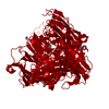
| Deposited unit | 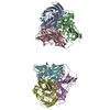
| ||||||||
|---|---|---|---|---|---|---|---|---|---|
| 1 | 
| ||||||||
| 2 | 
| ||||||||
| Unit cell |
| ||||||||
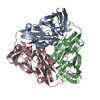
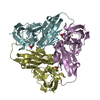
| Details | The physiological molecule is the homotrimer |
-Components
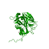
| #1: Protein | Mass: 35212.746 Da / Num. of mol.: 6 / Fragment: Residues 42-364, soluble domain Source method: isolated from a genetically manipulated source Source: (gene. exp.) Neisseria gonorrhoeae (bacteria) / Gene: aniA / Plasmid: pET / Production host: #2: Chemical | ChemComp-CU /   Mass: 63.546 Da / Num. of mol.: 12 / Source method: obtained synthetically / Formula: Cu Mass: 63.546 Da / Num. of mol.: 12 / Source method: obtained synthetically / Formula: Cu#3: Water | ChemComp-HOH / |  Mass: 18.015 Da / Num. of mol.: 1144 / Source method: isolated from a natural source / Formula: H2O Mass: 18.015 Da / Num. of mol.: 1144 / Source method: isolated from a natural source / Formula: H2O |
|---|
-Experimental details
-Experiment
| Experiment | Method: X-RAY DIFFRACTION / Number of used crystals: 1 |
|---|
- Sample preparation
| Crystal | Density Matthews: 2.97 Å3/Da / Density % sol: 58.55 % | ||||||||||||||||||||||||||||||||||||||||||
|---|---|---|---|---|---|---|---|---|---|---|---|---|---|---|---|---|---|---|---|---|---|---|---|---|---|---|---|---|---|---|---|---|---|---|---|---|---|---|---|---|---|---|---|
| Crystal grow | Temperature: 298 K / Method: vapor diffusion, hanging drop / pH: 10.5 Details: 1.7 M (NH4)2SO4, 0.2 M LiSO4 and 0.1 M CAPS, pH 10.5, VAPOR DIFFUSION, HANGING DROP, temperature 298K | ||||||||||||||||||||||||||||||||||||||||||
| Crystal grow | *PLUS Temperature: 22 ℃ / pH: 7 | ||||||||||||||||||||||||||||||||||||||||||
| Components of the solutions | *PLUS
|
-Data collection
| Diffraction | Mean temperature: 100 K |
|---|---|
| Diffraction source | Source: SYNCHROTRON / Site: Photon Factory  / Beamline: BL-6A / Wavelength: 1 Å / Beamline: BL-6A / Wavelength: 1 Å |
| Detector | Type: WEISSENBERG / Detector: DIFFRACTOMETER / Date: May 15, 1999 / Details: mirrors |
| Radiation | Monochromator: osmic mirrors / Protocol: SINGLE WAVELENGTH / Monochromatic (M) / Laue (L): M / Scattering type: x-ray |
| Radiation wavelength | Wavelength: 1 Å / Relative weight: 1 |
| Reflection | Resolution: 2.4→100 Å / Num. all: 95282 / Num. obs: 76914 / % possible obs: 80.7 % / Observed criterion σ(F): 2 / Observed criterion σ(I): 2 / Rmerge(I) obs: 0.073 / Net I/σ(I): 6.96 |
| Reflection shell | Resolution: 2.4→2.55 Å / Rmerge(I) obs: 0.227 / Mean I/σ(I) obs: 2.5 / Num. unique all: 11789 / % possible all: 74 |
| Reflection | *PLUS Highest resolution: 2.4 Å / Lowest resolution: 100 Å |
| Reflection shell | *PLUS Highest resolution: 2.4 Å / % possible obs: 74 % / Num. unique obs: 11789 / Rmerge(I) obs: 0.223 |
- Processing
| Software |
| |||||||||||||||||||||||||
|---|---|---|---|---|---|---|---|---|---|---|---|---|---|---|---|---|---|---|---|---|---|---|---|---|---|---|
| Refinement | Method to determine structure: MOLECULAR REPLACEMENT / Resolution: 2.4→50 Å / Isotropic thermal model: restrained / Cross valid method: THROUGHOUT / σ(F): 2 / Stereochemistry target values: Engh and Huber
| |||||||||||||||||||||||||
| Refinement step | Cycle: LAST / Resolution: 2.4→50 Å
| |||||||||||||||||||||||||
| Refine LS restraints |
| |||||||||||||||||||||||||
| LS refinement shell | Resolution: 2.4→2.55 Å / Num. reflection Rfree: 778 / Num. reflection obs: 11789 | |||||||||||||||||||||||||
| Software | *PLUS Name: CNS / Version: 1 / Classification: refinement | |||||||||||||||||||||||||
| Refinement | *PLUS Highest resolution: 2.4 Å / Lowest resolution: 50 Å / σ(F): 2 / % reflection Rfree: 10 % | |||||||||||||||||||||||||
| Solvent computation | *PLUS | |||||||||||||||||||||||||
| Displacement parameters | *PLUS Biso mean: 26.5 Å2 | |||||||||||||||||||||||||
| Refine LS restraints | *PLUS
| |||||||||||||||||||||||||
| LS refinement shell | *PLUS Highest resolution: 2.4 Å |
