 Movie
Movie Controller


+ Open data

- Basic information
| Entry | Database: PDB / ID: 1k95 | ||||||
|---|---|---|---|---|---|---|---|
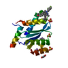
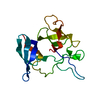
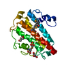
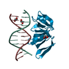
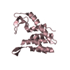
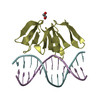
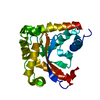
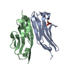
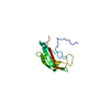
| Title | Crystal structure of des(1-52)grancalcin with bound calcium | ||||||
 Components Components | GRANCALCIN | ||||||
 Keywords Keywords | METAL BINDING PROTEIN / penta-EF-hand protein / calcium binding protein | ||||||
| Function / homology |  Function and homology information Function and homology informationazurophil granule lumen / membrane fusion / protein heterodimerization activity / calcium ion binding / Neutrophil degranulation / protein homodimerization activity / extracellular exosome / extracellular region / plasma membrane / cytoplasm / cytosol Similarity search - Function | ||||||
| Biological species |  Homo sapiens (human) Homo sapiens (human) | ||||||
| Method |  X-RAY DIFFRACTION / SYNCHROTRON / MOLECULAR REPLACEMENT / Resolution: 1.9 Å X-RAY DIFFRACTION / SYNCHROTRON / MOLECULAR REPLACEMENT / Resolution: 1.9 Å | ||||||
 Authors Authors | Jia, J. / Borregaard, N. / Lollike, K. / Cygler, M. | ||||||
 Citation Citation | Journal: Acta Crystallogr.,Sect.D / Year: 2001 Title: Structure of Ca(2+)-loaded human grancalcin. Authors: Jia, J. / Borregaard, N. / Lollike, K. / Cygler, M. #1: Journal: J.Mol.Biol. / Year: 2000Title: Crystal structure of human grancalcin, a member of the Penta-EF-Hand Protein family Authors: Jia, J. / Han, Q. / Borregaard, N. / Lollike, K. / Cygler, M. #2: Journal: Acta Crystallogr.,Sect.D / Year: 2000Title: Crystallizaiton and preliminary X-ray analysis of human grancalcin, a novel Ca2+-binding protein in leukocytes Authors: Han, Q. / Jia, J. / Li, Y. / Lollike, K. / Cygler, M. | ||||||
| History |
|
- Structure visualization
| Structure viewer | Molecule: MolmilJmol/JSmol |
|---|
- Downloads & links
-Download
| PDBx/mmCIF format | 1k95.cif.gz | 47.1 KB | Display | PDBx/mmCIF format |
|---|---|---|---|---|
| PDB format | pdb1k95.ent.gz | 33 KB | Display | PDB format |
| PDBx/mmJSON format | 1k95.json.gz | Tree view | PDBx/mmJSON format | |
| Others |  Other downloads Other downloads |
-Validation report
| Arichive directory | https://data.pdbj.org/pub/pdb/validation_reports/k9/1k95ftp://data.pdbj.org/pub/pdb/validation_reports/k9/1k95 | HTTPS FTP |
|---|
-Related structure data
| Related structure data | 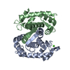 1k94C 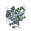 1f4qS S: Starting model for refinement C: citing same article ( |
|---|---|
| Similar structure data |

-Links
 PDBj
PDBj

- Assembly
| Deposited unit | 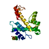
| ||||||||||
|---|---|---|---|---|---|---|---|---|---|---|---|
| 1 |
| ||||||||||
| 2 | 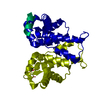
| ||||||||||
| Unit cell |
|
-Components
| #1: Protein | Mass: 18771.225 Da / Num. of mol.: 1 / Fragment: residues 53-217 Source method: isolated from a genetically manipulated source Source: (gene. exp.) Homo sapiens (human) / Production host:  |
|---|---|
| #2: Water | ChemComp-HOH /  Mass: 18.015 Da / Num. of mol.: 132 / Source method: isolated from a natural source / Formula: H2O Mass: 18.015 Da / Num. of mol.: 132 / Source method: isolated from a natural source / Formula: H2O |
-Experimental details
-Experiment
| Experiment | Method: X-RAY DIFFRACTION / Number of used crystals: 1 |
|---|
- Sample preparation
| Crystal | Density Matthews: 1.98 Å3/Da / Density % sol: 37.74 % | ||||||||||||||||||||||||||||||||||||
|---|---|---|---|---|---|---|---|---|---|---|---|---|---|---|---|---|---|---|---|---|---|---|---|---|---|---|---|---|---|---|---|---|---|---|---|---|---|
| Crystal grow | Temperature: 293 K / Method: vapor diffusion, hanging drop / pH: 4.5 Details: ammoniun sulfate, sodium acetate, pH 4.5, VAPOR DIFFUSION, HANGING DROP at 293K | ||||||||||||||||||||||||||||||||||||
| Crystal grow | *PLUS pH: 7.5 | ||||||||||||||||||||||||||||||||||||
| Components of the solutions | *PLUS
|
-Data collection
| Diffraction | Mean temperature: 100 K |
|---|---|
| Diffraction source | Source: SYNCHROTRON / Site: NSLS  / Beamline: X8C / Wavelength: 0.9795 Å / Beamline: X8C / Wavelength: 0.9795 Å |
| Detector | Type: ADSC QUANTUM 4 / Detector: CCD / Date: Jul 20, 2000 |
| Radiation | Protocol: SINGLE WAVELENGTH / Monochromatic (M) / Laue (L): M / Scattering type: x-ray |
| Radiation wavelength | Wavelength: 0.9795 Å / Relative weight: 1 |
| Reflection | Resolution: 1.9→50 Å / Num. all: 12769 / Num. obs: 12769 / % possible obs: 99.9 % / Redundancy: 12.8 % / Biso Wilson estimate: 17 Å2 / Rmerge(I) obs: 0.061 / Rsym value: 0.083 / Net I/σ(I): 13.5 |
| Reflection shell | Resolution: 1.9→1.94 Å / Rmerge(I) obs: 0.23 / Rsym value: 0.31 / % possible all: 99.3 |
| Reflection | *PLUS Lowest resolution: 50 Å / Num. measured all: 163585 |
| Reflection shell | *PLUS % possible obs: 99.3 % / Rmerge(I) obs: 0.23 |
- Processing
| Software |
| |||||||||||||||||||||||||
|---|---|---|---|---|---|---|---|---|---|---|---|---|---|---|---|---|---|---|---|---|---|---|---|---|---|---|
| Refinement | Method to determine structure: MOLECULAR REPLACEMENT Starting model: PDB ENTRY 1F4Q Resolution: 1.9→50 Å / Cross valid method: THROUGHOUT / σ(F): 0 / σ(I): 0 / Stereochemistry target values: Engh & Huber
| |||||||||||||||||||||||||
| Refinement step | Cycle: LAST / Resolution: 1.9→50 Å
| |||||||||||||||||||||||||
| Refine LS restraints |
| |||||||||||||||||||||||||
| Software | *PLUS Name: CNS / Classification: refinement | |||||||||||||||||||||||||
| Refinement | *PLUS Lowest resolution: 50 Å / σ(F): 0 / % reflection Rfree: 11.5 % / Rfactor obs: 0.211 | |||||||||||||||||||||||||
| Solvent computation | *PLUS | |||||||||||||||||||||||||
| Displacement parameters | *PLUS |
