 Movie
Movie Controller


+ Open data

- Basic information
| Entry | Database: PDB / ID: 1jxa | ||||||
|---|---|---|---|---|---|---|---|
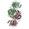
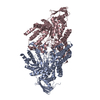
| Title | GLUCOSAMINE 6-PHOSPHATE SYNTHASE WITH GLUCOSE 6-PHOSPHATE | ||||||
 Components Components | glucosamine 6-phosphate synthase | ||||||
 Keywords Keywords | TRANSFERASE / beta-sandwich / nucleotide-binding fold / gene duplication / ammonia channel | ||||||
| Function / homology |  Function and homology information Function and homology informationglutamine-fructose-6-phosphate transaminase (isomerizing) / L-glutamine:D-fructose-6-phosphate transaminase (isomerizing) activity / UDP-N-acetylglucosamine metabolic process / UDP-N-acetylglucosamine biosynthetic process / carbohydrate derivative binding / fructose 6-phosphate metabolic process / protein N-linked glycosylation / carbohydrate metabolic process / cytosol Similarity search - Function | ||||||
| Biological species |  | ||||||
| Method |  X-RAY DIFFRACTION / SYNCHROTRON / MOLECULAR REPLACEMENT / Resolution: 3.1 Å X-RAY DIFFRACTION / SYNCHROTRON / MOLECULAR REPLACEMENT / Resolution: 3.1 Å | ||||||
 Authors Authors | Teplyakov, A. / Obmolova, G. / Badet, B. / Badet-Denisot, M.A. | ||||||
 Citation Citation | Journal: J.Mol.Biol. / Year: 2001 Title: Channeling of ammonia in glucosamine-6-phosphate synthase. Authors: Teplyakov, A. / Obmolova, G. / Badet, B. / Badet-Denisot, M.A. | ||||||
| History |
|
- Structure visualization
| Structure viewer | Molecule: MolmilJmol/JSmol |
|---|
- Downloads & links
-Download
| PDBx/mmCIF format | 1jxa.cif.gz | 354.9 KB | Display | PDBx/mmCIF format |
|---|---|---|---|---|
| PDB format | pdb1jxa.ent.gz | 287.9 KB | Display | PDB format |
| PDBx/mmJSON format | 1jxa.json.gz | Tree view | PDBx/mmJSON format | |
| Others |  Other downloads Other downloads |
-Validation report
| Arichive directory | https://data.pdbj.org/pub/pdb/validation_reports/jx/1jxaftp://data.pdbj.org/pub/pdb/validation_reports/jx/1jxa | HTTPS FTP |
|---|
-Related structure data
| Related structure data |  1gdo 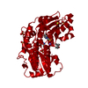 1moqS S: Starting model for refinement |
|---|---|
| Similar structure data |


-Links
 PDBj
PDBj


- Assembly
| Deposited unit | 
| ||||||||
|---|---|---|---|---|---|---|---|---|---|
| 1 | 
| ||||||||
| 2 | 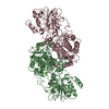
| ||||||||
| Unit cell |
| ||||||||
| Details | The second part of one biological homodimer is generated by the two-fold axis (-x, y, -z) applied to chain A. Another dimer is formed by chains B and C. |
-Components
| #1: Protein | Mass: 66818.000 Da / Num. of mol.: 3 Source method: isolated from a genetically manipulated source Source: (gene. exp.) References: UniProt: P17169, glutamine-fructose-6-phosphate transaminase (isomerizing) #2: Chemical |   Mass: 260.136 Da / Num. of mol.: 2 / Source method: obtained synthetically / Formula: C6H13O9P Mass: 260.136 Da / Num. of mol.: 2 / Source method: obtained synthetically / Formula: C6H13O9P#3: Water | ChemComp-HOH / |  Mass: 18.015 Da / Num. of mol.: 39 / Source method: isolated from a natural source / Formula: H2O Mass: 18.015 Da / Num. of mol.: 39 / Source method: isolated from a natural source / Formula: H2OHas protein modification | N | |
|---|
-Experimental details
-Experiment
| Experiment | Method: X-RAY DIFFRACTION / Number of used crystals: 1 |
|---|
- Sample preparation
| Crystal | Density Matthews: 3.39 Å3/Da / Density % sol: 63.69 % | ||||||||||||||||||||||||||||||||||||
|---|---|---|---|---|---|---|---|---|---|---|---|---|---|---|---|---|---|---|---|---|---|---|---|---|---|---|---|---|---|---|---|---|---|---|---|---|---|
| Crystal grow | Temperature: 295 K / Method: vapor diffusion, hanging drop / pH: 7 Details: LiCl, PEG4K, pH 7.0, VAPOR DIFFUSION, HANGING DROP, temperature 295K | ||||||||||||||||||||||||||||||||||||
| Crystal grow | *PLUS Temperature: 4 ℃ / Details: Obmolova, G., (1994) J.Mol.Biol., 242, 703. | ||||||||||||||||||||||||||||||||||||
| Components of the solutions | *PLUS
|
-Data collection
| Diffraction | Mean temperature: 100 K |
|---|---|
| Diffraction source | Source: SYNCHROTRON / Site: EMBL/DESY, HAMBURG  / Beamline: X11 / Wavelength: 0.9 Å / Beamline: X11 / Wavelength: 0.9 Å |
| Detector | Type: MARRESEARCH / Detector: IMAGE PLATE / Date: Apr 8, 1998 / Details: CYLINDRICAL MIRROR |
| Radiation | Monochromator: Ge(111) / Protocol: SINGLE WAVELENGTH / Monochromatic (M) / Laue (L): M / Scattering type: x-ray |
| Radiation wavelength | Wavelength: 0.9 Å / Relative weight: 1 |
| Reflection | Resolution: 3.1→20 Å / Num. all: 45685 / Num. obs: 45685 / % possible obs: 98.5 % / Observed criterion σ(F): 0 / Observed criterion σ(I): 0 / Redundancy: 2.4 % / Biso Wilson estimate: 74.5 Å2 / Rmerge(I) obs: 0.056 |
| Reflection shell | Resolution: 3.1→3.2 Å / Mean I/σ(I) obs: 2.8 / % possible all: 92 |
| Reflection | *PLUS Lowest resolution: 20 Å / Num. measured all: 111045 |
- Processing
| Software |
| |||||||||||||||||||||||||
|---|---|---|---|---|---|---|---|---|---|---|---|---|---|---|---|---|---|---|---|---|---|---|---|---|---|---|
| Refinement | Method to determine structure: MOLECULAR REPLACEMENT Starting model: 1MOQ, 1GDO Resolution: 3.1→12 Å / σ(F): 0 / Stereochemistry target values: Engh & Huber
| |||||||||||||||||||||||||
| Refinement step | Cycle: LAST / Resolution: 3.1→12 Å
| |||||||||||||||||||||||||
| Refine LS restraints |
| |||||||||||||||||||||||||
| Software | *PLUS Name: REFMAC / Classification: refinement | |||||||||||||||||||||||||
| Refinement | *PLUS Highest resolution: 3.1 Å / Lowest resolution: 12 Å / σ(F): 0 / % reflection Rfree: 5 % / Rfactor all: 0.22 / Rfactor obs: 0.2 / Rfactor Rfree: 0.28 / Rfactor Rwork: 0.2 | |||||||||||||||||||||||||
| Solvent computation | *PLUS | |||||||||||||||||||||||||
| Displacement parameters | *PLUS | |||||||||||||||||||||||||
| Refine LS restraints | *PLUS
|
