[English] 日本語
 Yorodumi
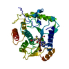
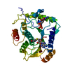
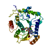
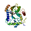
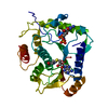
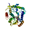
- PDB-1jtf: Crystal Structure Analysis of VP39-F180W mutant and m7GpppG complex -
+ Open data
- Basic information
| Entry | Database: PDB / ID: 1jtf | |||||||||
|---|---|---|---|---|---|---|---|---|---|---|
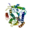
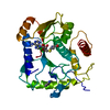
| Title | Crystal Structure Analysis of VP39-F180W mutant and m7GpppG complex | |||||||||
 Components Components | VP39 | |||||||||
 Keywords Keywords | TRANSFERASE / VP39 / mRNA Cap-binding protein / methyltransferase / mutant | |||||||||
| Function / homology |  Function and homology information Function and homology informationregulation of mRNA 3'-end processing / 7-methylguanosine mRNA capping / virion component / methyltransferase cap1 / methylation / methyltransferase cap1 activity / RNA binding Similarity search - Function | |||||||||
| Biological species |  Vaccinia virus Vaccinia virus | |||||||||
| Method |  X-RAY DIFFRACTION / X-RAY DIFFRACTION /  FOURIER SYNTHESIS / Resolution: 2.6 Å FOURIER SYNTHESIS / Resolution: 2.6 Å | |||||||||
 Authors Authors | Hu, G. / Oguro, A. / Gershon, P.D. / Quiocho, F.A. | |||||||||
 Citation Citation |  Journal: Biochemistry / Year: 2002 Journal: Biochemistry / Year: 2002Title: The "cap-binding slot" of an mRNA cap-binding protein: quantitative effects of aromatic side chain choice in the double-stacking sandwich with cap. Authors: Hu, G. / Oguro, A. / Li, C. / Gershon, P.D. / Quiocho, F.A. | |||||||||
| History |
|
- Structure visualization
| Structure viewer | Molecule:  Molmil Molmil Jmol/JSmol Jmol/JSmol |
|---|
- Downloads & links
- Download
| PDBx/mmCIF format |  1jtf.cif.gz 1jtf.cif.gz | 72.7 KB | Display |  PDBx/mmCIF format PDBx/mmCIF format |
|---|---|---|---|---|
| PDB format |  pdb1jtf.ent.gz pdb1jtf.ent.gz | 53 KB | Display |  PDB format PDB format |
| PDBx/mmJSON format |  1jtf.json.gz 1jtf.json.gz | Tree view |  PDBx/mmJSON format PDBx/mmJSON format | |
| Others |  Other downloads Other downloads |
-Validation report
| Summary document |  1jtf_validation.pdf.gz 1jtf_validation.pdf.gz | 1 MB | Display |  wwPDB validaton report wwPDB validaton report |
|---|---|---|---|---|
| Full document |  1jtf_full_validation.pdf.gz 1jtf_full_validation.pdf.gz | 1 MB | Display | |
| Data in XML |  1jtf_validation.xml.gz 1jtf_validation.xml.gz | 14.8 KB | Display | |
| Data in CIF |  1jtf_validation.cif.gz 1jtf_validation.cif.gz | 19.8 KB | Display | |
| Arichive directory |  https://data.pdbj.org/pub/pdb/validation_reports/jt/1jtf https://data.pdbj.org/pub/pdb/validation_reports/jt/1jtf ftp://data.pdbj.org/pub/pdb/validation_reports/jt/1jtf ftp://data.pdbj.org/pub/pdb/validation_reports/jt/1jtf | HTTPS FTP |
-Related structure data
- Links
Links
- Assembly
| Deposited unit | 
| ||||||||
|---|---|---|---|---|---|---|---|---|---|
| 1 |
| ||||||||
| Unit cell |
|
- Components
| #1: Protein | Mass: 35960.473 Da / Num. of mol.: 1 / Fragment: residues 1-307 / Mutation: F180W Source method: isolated from a genetically manipulated source Source: (gene. exp.)  Vaccinia virus / Genus: Orthopoxvirus / Plasmid: pPG177 / Production host: Vaccinia virus / Genus: Orthopoxvirus / Plasmid: pPG177 / Production host:  References: UniProt: P07617, polynucleotide adenylyltransferase |
|---|---|
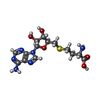
| #2: Chemical | ChemComp-SAH / |
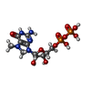
| #3: Chemical | ChemComp-M7G / |
| #4: Water | ChemComp-HOH / |
-Experimental details
-Experiment
| Experiment | Method:  X-RAY DIFFRACTION / Number of used crystals: 1 X-RAY DIFFRACTION / Number of used crystals: 1 |
|---|
- Sample preparation
| Crystal | Density Matthews: 2.79 Å3/Da / Density % sol: 55.84 % | ||||||||||||||||||||||||
|---|---|---|---|---|---|---|---|---|---|---|---|---|---|---|---|---|---|---|---|---|---|---|---|---|---|
| Crystal grow | Temperature: 298 K / Method: vapor diffusion, hanging drop / pH: 4.5 Details: PEG 8000, sodium citrate, ammonium sulphate, pH 4.5, VAPOR DIFFUSION, HANGING DROP, temperature 298K | ||||||||||||||||||||||||
| Crystal grow | *PLUS Method: unknownDetails: Hodel, A.E., (1996) Cell(Cambridge,Mass.), 85, 247. | ||||||||||||||||||||||||
| Components of the solutions | *PLUS
|
-Data collection
| Diffraction | Mean temperature: 113 K |
|---|---|
| Diffraction source | Source:  ROTATING ANODE / Type: RIGAKU RU200 / Wavelength: 1.5418 Å ROTATING ANODE / Type: RIGAKU RU200 / Wavelength: 1.5418 Å |
| Detector | Type: SIEMENS / Detector: CCD / Date: May 13, 1999 |
| Radiation | Monochromator: Graphite / Protocol: SINGLE WAVELENGTH / Monochromatic (M) / Laue (L): M / Scattering type: x-ray |
| Radiation wavelength | Wavelength: 1.5418 Å / Relative weight: 1 |
| Reflection | Resolution: 2.6→50 Å / Num. all: 22968 / Num. obs: 21940 / % possible obs: 94.1 % / Observed criterion σ(F): 2 / Observed criterion σ(I): 2 |
| Reflection shell | Resolution: 2.61→2.74 Å / % possible all: 50 |
| Reflection | *PLUS Rmerge(I) obs: 0.107 |
- Processing
| Software |
| |||||||||||||||||||||||||
|---|---|---|---|---|---|---|---|---|---|---|---|---|---|---|---|---|---|---|---|---|---|---|---|---|---|---|
| Refinement | Method to determine structure:  FOURIER SYNTHESIS / Resolution: 2.6→8 Å / σ(F): 2 / Stereochemistry target values: Engh & Huber FOURIER SYNTHESIS / Resolution: 2.6→8 Å / σ(F): 2 / Stereochemistry target values: Engh & Huber
| |||||||||||||||||||||||||
| Refinement step | Cycle: LAST / Resolution: 2.6→8 Å
| |||||||||||||||||||||||||
| Refine LS restraints |
| |||||||||||||||||||||||||
| LS refinement shell | Resolution: 2.61→2.74 Å / Rfactor Rfree error: 0.008
| |||||||||||||||||||||||||
| Refinement | *PLUS Rfactor obs: 0.273 / Rfactor Rfree: 0.279 / Rfactor Rwork: 0.258 | |||||||||||||||||||||||||
| Solvent computation | *PLUS | |||||||||||||||||||||||||
| Displacement parameters | *PLUS | |||||||||||||||||||||||||
| Refine LS restraints | *PLUS
| |||||||||||||||||||||||||
| LS refinement shell | *PLUS Rfactor Rfree: 0.3 / Rfactor Rwork: 0.27 |
 Movie
Movie Controller






 PDBj
PDBj
