 Movie
Movie Controller


+ Open data

- Basic information
| Entry | Database: PDB / ID: 1jme | ||||||
|---|---|---|---|---|---|---|---|
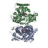
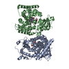
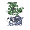
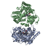
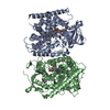
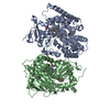
| Title | Crystal Structure of Phe393His Cytochrome P450 BM3 | ||||||
 Components Components | BIFUNCTIONAL P-450:NADPH-P450 REDUCTASE | ||||||
 Keywords Keywords | OXIDOREDUCTASE / CYTOCHROME P450 / FATTY ACID HYDROXYLASE / MONOOXYGENASE | ||||||
| Function / homology |  Function and homology information Function and homology informationaromatase activity / NADPH-hemoprotein reductase / NADPH-hemoprotein reductase activity / oxidoreductase activity, acting on paired donors, with incorporation or reduction of molecular oxygen, reduced flavin or flavoprotein as one donor, and incorporation of one atom of oxygen / unspecific monooxygenase / FMN binding / flavin adenine dinucleotide binding / iron ion binding / heme binding / identical protein binding / cytosol Similarity search - Function | ||||||
| Biological species |  Bacillus megaterium (bacteria) Bacillus megaterium (bacteria) | ||||||
| Method |  X-RAY DIFFRACTION / SYNCHROTRON / MOLECULAR REPLACEMENT / Resolution: 2 Å X-RAY DIFFRACTION / SYNCHROTRON / MOLECULAR REPLACEMENT / Resolution: 2 Å | ||||||
 Authors Authors | Ost, T.W.B. / Munro, A.W. / Mowat, C.G. / Pesseguiero, A. / Fulco, A.J. / Cho, A.K. / Cheesman, M.A. / Walkinshaw, M.D. / Chapman, S.K. | ||||||
 Citation Citation | Journal: Biochemistry / Year: 2001 Title: Structural and spectroscopic analysis of the F393H mutant of flavocytochrome P450 BM3. Authors: Ost, T.W. / Munro, A.W. / Mowat, C.G. / Taylor, P.R. / Pesseguiero, A. / Fulco, A.J. / Cho, A.K. / Cheesman, M.A. / Walkinshaw, M.D. / Chapman, S.K. | ||||||
| History |
|
- Structure visualization
| Structure viewer | Molecule: MolmilJmol/JSmol |
|---|
- Downloads & links
-Download
| PDBx/mmCIF format | 1jme.cif.gz | 215.5 KB | Display | PDBx/mmCIF format |
|---|---|---|---|---|
| PDB format | pdb1jme.ent.gz | 169.2 KB | Display | PDB format |
| PDBx/mmJSON format | 1jme.json.gz | Tree view | PDBx/mmJSON format | |
| Others |  Other downloads Other downloads |
-Validation report
| Summary document | 1jme_validation.pdf.gz | 547.1 KB | Display | wwPDB validaton report |
|---|---|---|---|---|
| Full document | 1jme_full_validation.pdf.gz | 561.9 KB | Display | |
| Data in XML | 1jme_validation.xml.gz | 20 KB | Display | |
| Data in CIF | 1jme_validation.cif.gz | 36.3 KB | Display | |
| Arichive directory | https://data.pdbj.org/pub/pdb/validation_reports/jm/1jmeftp://data.pdbj.org/pub/pdb/validation_reports/jm/1jme | HTTPS FTP |
-Related structure data
| Related structure data |  2hpdS S: Starting model for refinement |
|---|---|
| Similar structure data |


-Links
 PDBj
PDBj


- Assembly
| Deposited unit | 
| ||||||||||
|---|---|---|---|---|---|---|---|---|---|---|---|
| 1 |
| ||||||||||
| Unit cell |
|
-Components
| #1: Protein | Mass: 52160.434 Da / Num. of mol.: 2 / Fragment: CYTOCHROME P450 / Mutation: F393H Source method: isolated from a genetically manipulated source Source: (gene. exp.) Bacillus megaterium (bacteria) / Gene: CYP102A1 / Plasmid: pCM81 / Production host: #2: Chemical |   Mass: 616.487 Da / Num. of mol.: 2 / Source method: obtained synthetically / Formula: C34H32FeN4O4 Mass: 616.487 Da / Num. of mol.: 2 / Source method: obtained synthetically / Formula: C34H32FeN4O4#3: Water | ChemComp-HOH / |  Mass: 18.015 Da / Num. of mol.: 1182 / Source method: isolated from a natural source / Formula: H2O Mass: 18.015 Da / Num. of mol.: 1182 / Source method: isolated from a natural source / Formula: H2O |
|---|
-Experimental details
-Experiment
| Experiment | Method: X-RAY DIFFRACTION / Number of used crystals: 1 |
|---|
- Sample preparation
| Crystal | Density Matthews: 2.64 Å3/Da / Density % sol: 53.49 % | ||||||||||||||||||||||||||||||||||||||||||
|---|---|---|---|---|---|---|---|---|---|---|---|---|---|---|---|---|---|---|---|---|---|---|---|---|---|---|---|---|---|---|---|---|---|---|---|---|---|---|---|---|---|---|---|
| Crystal grow | Temperature: 277 K / Method: vapor diffusion, hanging drop / pH: 6.5 Details: PEG 8000, PIPES, magnesium sulfate, pH 6.5, VAPOR DIFFUSION, HANGING DROP at 277K | ||||||||||||||||||||||||||||||||||||||||||
| Crystal grow | *PLUS Temperature: 4 ℃ / pH: 7.4 | ||||||||||||||||||||||||||||||||||||||||||
| Components of the solutions | *PLUS
|
-Data collection
| Diffraction | Mean temperature: 100 K |
|---|---|
| Diffraction source | Source: SYNCHROTRON / Site: SRS  / Beamline: PX9.5 / Wavelength: 1.3 Å / Beamline: PX9.5 / Wavelength: 1.3 Å |
| Detector | Type: ADSC QUANTUM 4 / Detector: CCD / Date: May 20, 2000 / Details: mirrors |
| Radiation | Protocol: SINGLE WAVELENGTH / Monochromatic (M) / Laue (L): M / Scattering type: x-ray |
| Radiation wavelength | Wavelength: 1.3 Å / Relative weight: 1 |
| Reflection | Resolution: 2→40 Å / Num. all: 73016 / Num. obs: 73016 / % possible obs: 96.9 % / Observed criterion σ(F): 0 / Observed criterion σ(I): 0 / Redundancy: 6.58 % / Biso Wilson estimate: 25.5 Å2 / Rmerge(I) obs: 0.054 / Net I/σ(I): 19.4 |
| Reflection shell | Resolution: 2→2.07 Å |
| Reflection | *PLUS Num. measured all: 480222 |
| Reflection shell | *PLUS Rmerge(I) obs: 0.138 |
- Processing
| Software |
| |||||||||||||||||||||||||
|---|---|---|---|---|---|---|---|---|---|---|---|---|---|---|---|---|---|---|---|---|---|---|---|---|---|---|
| Refinement | Method to determine structure: MOLECULAR REPLACEMENT Starting model: PDB ENTRY 2HPD Resolution: 2→40 Å / Cross valid method: THROUGHOUT
| |||||||||||||||||||||||||
| Refinement step | Cycle: LAST / Resolution: 2→40 Å
| |||||||||||||||||||||||||
| Refine LS restraints |
| |||||||||||||||||||||||||
| Software | *PLUS Name: REFMAC / Classification: refinement | |||||||||||||||||||||||||
| Refinement | *PLUS Highest resolution: 2 Å / % reflection Rfree: 5.2 % | |||||||||||||||||||||||||
| Solvent computation | *PLUS | |||||||||||||||||||||||||
| Displacement parameters | *PLUS | |||||||||||||||||||||||||
| Refine LS restraints | *PLUS Type: p_angle_d / Dev ideal: 1.8 |
