 Movie
Movie Controller


+ Open data

- Basic information
| Entry | Database: PDB / ID: 1jh5 | ||||||
|---|---|---|---|---|---|---|---|
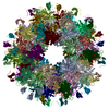
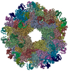
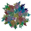
| Title | Crystal Structure of sTALL-1 of TNF family ligand | ||||||
 Components Components | TUMOR NECROSIS FACTOR LIGAND SUPERFAMILY MEMBER 13B | ||||||
 Keywords Keywords | IMMUNE SYSTEM / ANTITUMOR PROTEIN / TALL-1 / BLYS / THANK / BAFF | ||||||
| Function / homology |  Function and homology information Function and homology informationB cell costimulation / positive regulation of germinal center formation / lymphocyte homeostasis / TNFs bind their physiological receptors / TNF receptor superfamily (TNFSF) members mediating non-canonical NF-kB pathway / transitional one stage B cell differentiation / germinal center formation / tumor necrosis factor receptor binding / skin development / B cell homeostasis ...B cell costimulation / positive regulation of germinal center formation / lymphocyte homeostasis / TNFs bind their physiological receptors / TNF receptor superfamily (TNFSF) members mediating non-canonical NF-kB pathway / transitional one stage B cell differentiation / germinal center formation / tumor necrosis factor receptor binding / skin development / B cell homeostasis / B cell proliferation / T cell proliferation / T cell costimulation / positive regulation of B cell proliferation / positive regulation of T cell proliferation / cytokine activity / tumor necrosis factor-mediated signaling pathway / TNFR2 non-canonical NF-kB pathway / immune response / signaling receptor binding / focal adhesion / perinuclear region of cytoplasm / signal transduction / : / extracellular region / membrane / plasma membrane / cytoplasm Similarity search - Function | ||||||
| Biological species |  Homo sapiens (human) Homo sapiens (human) | ||||||
| Method |  X-RAY DIFFRACTION / Resolution: 3 Å X-RAY DIFFRACTION / Resolution: 3 Å | ||||||
 Authors Authors | Liu, Y. / Xu, L. / Opalka, N. / Shu, H.-B. / Zhang, G. | ||||||
 Citation Citation | Journal: Cell(Cambridge,Mass.) / Year: 2002 Title: Crystal structure of sTALL-1 reveals a virus-like assembly of TNF family ligands. Authors: Liu, Y. / Xu, L. / Opalka, N. / Kappler, J. / Shu, H.B. / Zhang, G. | ||||||
| History |
|
- Structure visualization
| Structure viewer | Molecule: MolmilJmol/JSmol |
|---|
- Downloads & links
-Download
| PDBx/mmCIF format | 1jh5.cif.gz | 276.9 KB | Display | PDBx/mmCIF format |
|---|---|---|---|---|
| PDB format | pdb1jh5.ent.gz | 228.2 KB | Display | PDB format |
| PDBx/mmJSON format | 1jh5.json.gz | Tree view | PDBx/mmJSON format | |
| Others |  Other downloads Other downloads |
-Validation report
| Arichive directory | https://data.pdbj.org/pub/pdb/validation_reports/jh/1jh5ftp://data.pdbj.org/pub/pdb/validation_reports/jh/1jh5 | HTTPS FTP |
|---|
-Related structure data
| Similar structure data |
|---|
-Links
 PDBj
PDBj- Assembly
| Deposited unit | 
| ||||||||||
|---|---|---|---|---|---|---|---|---|---|---|---|
| 1 | x 6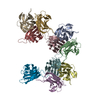
| ||||||||||
| Unit cell |
|
-Components
| #1: Protein | Mass: 16244.602 Da / Num. of mol.: 10 / Fragment: sTALL-1, soluble part of TALL-1 Source method: isolated from a genetically manipulated source Source: (gene. exp.) Homo sapiens (human) / Production host:  |
|---|
-Experimental details
-Experiment
| Experiment | Method: X-RAY DIFFRACTION / Number of used crystals: 3 |
|---|
- Sample preparation
| Crystal | Density Matthews: 5.18 Å3/Da / Density % sol: 76.26 % | ||||||||||||||||||||||||||||||||||||||||||
|---|---|---|---|---|---|---|---|---|---|---|---|---|---|---|---|---|---|---|---|---|---|---|---|---|---|---|---|---|---|---|---|---|---|---|---|---|---|---|---|---|---|---|---|
| Crystal grow | Temperature: 277 K / Method: vapor diffusion, hanging drop / pH: 9 Details: 35% dioxane, pH 9.0, VAPOR DIFFUSION, HANGING DROP at 277K | ||||||||||||||||||||||||||||||||||||||||||
| Crystal grow | *PLUS Method: vapor diffusion | ||||||||||||||||||||||||||||||||||||||||||
| Components of the solutions | *PLUS
|
-Data collection
| Diffraction | Mean temperature: 93 K |
|---|---|
| Diffraction source | Source: ROTATING ANODE / Type: RIGAKU RU200 / Wavelength: 1.5418 |
| Detector | Type: RIGAKU RAXIS IV / Detector: IMAGE PLATE |
| Radiation | Protocol: SINGLE WAVELENGTH / Monochromatic (M) / Laue (L): M / Scattering type: x-ray |
| Radiation wavelength | Wavelength: 1.5418 Å / Relative weight: 1 |
| Reflection | Resolution: 3→20 Å / Num. all: 69018 / Num. obs: 66001 / % possible obs: 95.7 % / Observed criterion σ(F): 0 / Observed criterion σ(I): 0 / Redundancy: 5 % / Rmerge(I) obs: 0.117 / Net I/σ(I): 12 |
| Reflection | *PLUS Num. measured all: 334821 |
- Processing
| Software |
| ||||||||||||||||||||
|---|---|---|---|---|---|---|---|---|---|---|---|---|---|---|---|---|---|---|---|---|---|
| Refinement | Resolution: 3→19.99 Å / Rfactor Rfree error: 0.005 / Data cutoff high absF: 172269.28 / Data cutoff low absF: 0 / Isotropic thermal model: RESTRAINED / Cross valid method: THROUGHOUT / σ(F): 2
| ||||||||||||||||||||
| Solvent computation | Solvent model: FLAT MODEL / Bsol: 10 Å2 / ksol: 0.285 e/Å3 | ||||||||||||||||||||
| Displacement parameters | Biso mean: 30.6 Å2
| ||||||||||||||||||||
| Refine analyze |
| ||||||||||||||||||||
| Refinement step | Cycle: LAST / Resolution: 3→19.99 Å
| ||||||||||||||||||||
| Refine LS restraints |
| ||||||||||||||||||||
| LS refinement shell | Resolution: 3→3.19 Å / Rfactor Rfree error: 0.021 / Total num. of bins used: 6
| ||||||||||||||||||||
| Xplor file |
| ||||||||||||||||||||
| Software | *PLUS Name: CNS / Version: 1 / Classification: refinement | ||||||||||||||||||||
| Refinement | *PLUS Num. reflection obs: 51752 / % reflection Rfree: 5 % | ||||||||||||||||||||
| Solvent computation | *PLUS | ||||||||||||||||||||
| Displacement parameters | *PLUS Biso mean: 30.6 Å2 | ||||||||||||||||||||
| Refine LS restraints | *PLUS
| ||||||||||||||||||||
| LS refinement shell | *PLUS Rfactor Rfree: 0.344 / % reflection Rfree: 5 % / Rfactor Rwork: 0.331 |
