 Movie
Movie Controller


+ Open data

- Basic information
| Entry | Database: PDB / ID: 1jec | ||||||
|---|---|---|---|---|---|---|---|
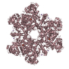
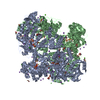
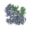
| Title | Crystal Structure of ATP Sulfurylase in complex with thiosulfate | ||||||
 Components Components | SULFATE ADENYLYLTRANSFERASE | ||||||
 Keywords Keywords | TRANSFERASE / alpha-beta protein / beta-barrel / Rossmann-fold / inhibitor complex / thiosulfate | ||||||
| Function / homology |  Function and homology information Function and homology information: / sulfur amino acid metabolic process / sulfate adenylyltransferase / sulfate adenylyltransferase (ATP) activity / hydrogen sulfide biosynthetic process / mitochondrion / ATP binding / cytoplasm Similarity search - Function | ||||||
| Biological species |  | ||||||
| Method |  X-RAY DIFFRACTION / MOLECULAR REPLACEMENT / Resolution: 2.5 Å X-RAY DIFFRACTION / MOLECULAR REPLACEMENT / Resolution: 2.5 Å | ||||||
 Authors Authors | Ullrich, T.C. / Huber, R. | ||||||
 Citation Citation | Journal: J.Mol.Biol. / Year: 2001 Title: The complex structures of ATP sulfurylase with thiosulfate, ADP and chlorate reveal new insights in inhibitory effects and the catalytic cycle. Authors: Ullrich, T.C. / Huber, R. | ||||||
| History |
|
- Structure visualization
| Structure viewer | Molecule: MolmilJmol/JSmol |
|---|
- Downloads & links
-Download
| PDBx/mmCIF format | 1jec.cif.gz | 131.5 KB | Display | PDBx/mmCIF format |
|---|---|---|---|---|
| PDB format | pdb1jec.ent.gz | 99.5 KB | Display | PDB format |
| PDBx/mmJSON format | 1jec.json.gz | Tree view | PDBx/mmJSON format | |
| Others |  Other downloads Other downloads |
-Validation report
| Arichive directory | https://data.pdbj.org/pub/pdb/validation_reports/je/1jecftp://data.pdbj.org/pub/pdb/validation_reports/je/1jec | HTTPS FTP |
|---|
-Related structure data
| Related structure data |  1jedC  1jeeC  1g8fS S: Starting model for refinement C: citing same article ( |
|---|---|
| Similar structure data |


-Links
 PDBj
PDBj

- Assembly
| Deposited unit | 
| ||||||||
|---|---|---|---|---|---|---|---|---|---|
| 1 | x 6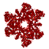
| ||||||||
| Unit cell |
| ||||||||
| Components on special symmetry positions |
| ||||||||
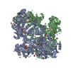
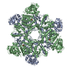
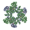
| Details | Homohexamer, generated by the D3 symmetry of the R32 spacegroup (1 subunit per asymmetric unit) |
-Components
-Protein , 1 types, 1 molecules A
| #1: Protein | Mass: 57661.043 Da / Num. of mol.: 1 / Source method: isolated from a natural source / Details: Native purification out of yeast cells / Source: (natural) |
|---|
-Non-polymers , 8 types, 466 molecules 


| #2: Chemical | ChemComp-CD /  Mass: 112.411 Da / Num. of mol.: 7 / Source method: obtained synthetically / Formula: Cd Mass: 112.411 Da / Num. of mol.: 7 / Source method: obtained synthetically / Formula: Cd#3: Chemical |  Mass: 40.078 Da / Num. of mol.: 3 / Source method: obtained synthetically / Formula: Ca Mass: 40.078 Da / Num. of mol.: 3 / Source method: obtained synthetically / Formula: Ca#4: Chemical | ChemComp-MG / |  Mass: 24.305 Da / Num. of mol.: 1 / Source method: obtained synthetically / Formula: Mg Mass: 24.305 Da / Num. of mol.: 1 / Source method: obtained synthetically / Formula: Mg#5: Chemical |  Mass: 22.990 Da / Num. of mol.: 3 / Source method: obtained synthetically / Formula: Na Mass: 22.990 Da / Num. of mol.: 3 / Source method: obtained synthetically / Formula: Na#6: Chemical | ChemComp-SO4 / |  Mass: 96.063 Da / Num. of mol.: 1 / Source method: obtained synthetically / Formula: SO4 Mass: 96.063 Da / Num. of mol.: 1 / Source method: obtained synthetically / Formula: SO4#7: Chemical | ChemComp-THJ / |  Mass: 112.128 Da / Num. of mol.: 1 / Source method: obtained synthetically / Formula: O3S2 Mass: 112.128 Da / Num. of mol.: 1 / Source method: obtained synthetically / Formula: O3S2#8: Chemical | ChemComp-ACY /  Mass: 60.052 Da / Num. of mol.: 12 / Source method: obtained synthetically / Formula: C2H4O2 Mass: 60.052 Da / Num. of mol.: 12 / Source method: obtained synthetically / Formula: C2H4O2#9: Water | ChemComp-HOH / | Mass: 18.015 Da / Num. of mol.: 438 / Source method: isolated from a natural source / Formula: H2O |
|---|
-Experimental details
-Experiment
| Experiment | Method: X-RAY DIFFRACTION / Number of used crystals: 1 |
|---|
- Sample preparation
| Crystal | Density Matthews: 3.38 Å3/Da / Density % sol: 64 % | ||||||||||||||||||||||||||||||||||||||||||||||||
|---|---|---|---|---|---|---|---|---|---|---|---|---|---|---|---|---|---|---|---|---|---|---|---|---|---|---|---|---|---|---|---|---|---|---|---|---|---|---|---|---|---|---|---|---|---|---|---|---|---|
| Crystal grow | Temperature: 291 K / Method: vapor diffusion, hanging drop / pH: 7.5 Details: 500 mM NaAc, 50 mM HEPES, 25 mM CdSO4, pH 7.5, VAPOR DIFFUSION, HANGING DROP, temperature 291K | ||||||||||||||||||||||||||||||||||||||||||||||||
| Crystal grow | *PLUS Temperature: 18 ℃ / Details: Ullrich, T.C., (2001) EMBO J., 20, 316. | ||||||||||||||||||||||||||||||||||||||||||||||||
| Components of the solutions | *PLUS
|
-Data collection
| Diffraction | Mean temperature: 100 K |
|---|---|
| Diffraction source | Source: ROTATING ANODE / Type: RIGAKU RU200 / Wavelength: 1.542 Å |
| Detector | Type: MARRESEARCH / Detector: IMAGE PLATE / Date: Jan 10, 2001 / Details: mirrors |
| Radiation | Monochromator: graphite / Protocol: SINGLE WAVELENGTH / Monochromatic (M) / Laue (L): M / Scattering type: x-ray |
| Radiation wavelength | Wavelength: 1.542 Å / Relative weight: 1 |
| Reflection | Resolution: 2.5→25 Å / Num. all: 26847 / Num. obs: 26794 / % possible obs: 99.8 % / Observed criterion σ(F): 0 / Observed criterion σ(I): 2 / Redundancy: 5.5 % / Biso Wilson estimate: 42.2 Å2 / Rmerge(I) obs: 0.09 / Rsym value: 0.09 / Net I/σ(I): 7.6 |
| Reflection shell | Resolution: 2.5→2.66 Å / Redundancy: 5.5 % / Rmerge(I) obs: 0.437 / Mean I/σ(I) obs: 1.7 / Num. unique all: 4187 / Rsym value: 0.437 / % possible all: 99.5 |
| Reflection | *PLUS Rmerge(I) obs: 0.09 |
| Reflection shell | *PLUS % possible obs: 99.5 % |
- Processing
| Software |
| ||||||||||||||||||||||||||||||||||||||||||||
|---|---|---|---|---|---|---|---|---|---|---|---|---|---|---|---|---|---|---|---|---|---|---|---|---|---|---|---|---|---|---|---|---|---|---|---|---|---|---|---|---|---|---|---|---|---|
| Refinement | Method to determine structure: MOLECULAR REPLACEMENT Starting model: 1G8F Resolution: 2.5→24.34 Å / Rfactor Rfree error: 0.006 / Data cutoff high absF: 2907948.18 / Data cutoff low absF: 0 / Isotropic thermal model: RESTRAINED / Cross valid method: THROUGHOUT / σ(F): 0 / σ(I): 0 / Stereochemistry target values: Engh & Huber
| ||||||||||||||||||||||||||||||||||||||||||||
| Solvent computation | Solvent model: FLAT MODEL / Bsol: 41.4 Å2 / ksol: 0.336 e/Å3 | ||||||||||||||||||||||||||||||||||||||||||||
| Displacement parameters | Biso mean: 34.2 Å2
| ||||||||||||||||||||||||||||||||||||||||||||
| Refine analyze |
| ||||||||||||||||||||||||||||||||||||||||||||
| Refinement step | Cycle: LAST / Resolution: 2.5→24.34 Å
| ||||||||||||||||||||||||||||||||||||||||||||
| Refine LS restraints |
| ||||||||||||||||||||||||||||||||||||||||||||
| LS refinement shell | Resolution: 2.5→2.66 Å / Rfactor Rfree error: 0.02 / Total num. of bins used: 6
| ||||||||||||||||||||||||||||||||||||||||||||
| Software | *PLUS Name: CNS / Version: 1 / Classification: refinement | ||||||||||||||||||||||||||||||||||||||||||||
| Refinement | *PLUS σ(F): 0 / % reflection Rfree: 5.2 % | ||||||||||||||||||||||||||||||||||||||||||||
| Solvent computation | *PLUS | ||||||||||||||||||||||||||||||||||||||||||||
| Displacement parameters | *PLUS Biso mean: 34.2 Å2 | ||||||||||||||||||||||||||||||||||||||||||||
| Refine LS restraints | *PLUS
| ||||||||||||||||||||||||||||||||||||||||||||
| LS refinement shell | *PLUS Rfactor Rfree: 0.299 / % reflection Rfree: 5.3 % / Rfactor Rwork: 0.227 |
