 Movie
Movie Controller

[English] 日本語
 Yorodumi
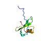
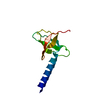
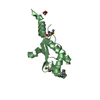
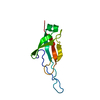
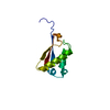
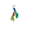
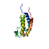
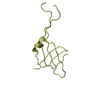
- PDB-1je4: Solution structure of the monomeric variant of the chemokine MIP-1beta -
+ Open data

- Basic information
| Entry | Database: PDB / ID: 1je4 | ||||||
|---|---|---|---|---|---|---|---|
| Title | Solution structure of the monomeric variant of the chemokine MIP-1beta | ||||||
 Components Components | macrophage inflammatory protein 1-beta | ||||||
 Keywords Keywords | ANTIVIRAL PROTEIN / MIP-1beta / chemokine / macrophage inflammatory protein | ||||||
| Function / homology |  Function and homology information Function and homology informationCCR1 chemokine receptor binding / positive regulation of natural killer cell chemotaxis / CCR5 chemokine receptor binding / CCR chemokine receptor binding / positive regulation of calcium ion transport / eosinophil chemotaxis / chemokine-mediated signaling pathway / Chemokine receptors bind chemokines / chemokine activity / establishment or maintenance of cell polarity ...CCR1 chemokine receptor binding / positive regulation of natural killer cell chemotaxis / CCR5 chemokine receptor binding / CCR chemokine receptor binding / positive regulation of calcium ion transport / eosinophil chemotaxis / chemokine-mediated signaling pathway / Chemokine receptors bind chemokines / chemokine activity / establishment or maintenance of cell polarity / Interleukin-10 signaling / negative regulation by host of viral transcription / positive regulation of calcium-mediated signaling / cytokine activity / response to virus / response to toxic substance / antimicrobial humoral immune response mediated by antimicrobial peptide / cell-cell signaling / G alpha (i) signalling events / cell adhesion / positive regulation of cell migration / inflammatory response / immune response / signal transduction / extracellular space / extracellular region / identical protein binding Similarity search - Function | ||||||
| Biological species |  Homo sapiens (human) Homo sapiens (human) | ||||||
| Method | SOLUTION NMR / The initial fold was obtained by distance geometry, further refined by simulated annealing. | ||||||
| Model type details | minimized average | ||||||
 Authors Authors | Kim, S. / Jao, S. / Laurence, J.S. / LiWang, P.J. | ||||||
 Citation Citation | Journal: Biochemistry / Year: 2001 Title: Structural comparison of monomeric variants of the chemokine MIP-1beta having differing ability to bind the receptor CCR5. Authors: Kim, S. / Jao, S. / Laurence, J.S. / LiWang, P.J. #1: Journal: Biochemistry / Year: 2000Title: CC chemokine MIP-1beta can function as a monomer and depends on Phe13 for receptor binding Authors: Laurence, J.S. / Blanpain, C. / Burgner, J.W. / Parmentier, M. / LiWang, P.J. #2: Journal: Science / Year: 1994Title: High-resolution solution structure of the beta chemokine hMIP-1beta by multidimensional NMR Authors: Lodi, P.J. / Garrett, D.S. / Kuszewski, J. / Tsang, M.L. / Weatherbee, J.A. / Leonard, W.J. / Gronenborn, A.M. / Clore, G.M. | ||||||
| History |
|
- Structure visualization
| Structure viewer | Molecule: MolmilJmol/JSmol |
|---|
- Downloads & links
-Download
| PDBx/mmCIF format | 1je4.cif.gz | 30.2 KB | Display | PDBx/mmCIF format |
|---|---|---|---|---|
| PDB format | pdb1je4.ent.gz | 23.4 KB | Display | PDB format |
| PDBx/mmJSON format | 1je4.json.gz | Tree view | PDBx/mmJSON format | |
| Others |  Other downloads Other downloads |
-Validation report
| Summary document | 1je4_validation.pdf.gz | 243.1 KB | Display | wwPDB validaton report |
|---|---|---|---|---|
| Full document | 1je4_full_validation.pdf.gz | 242.8 KB | Display | |
| Data in XML | 1je4_validation.xml.gz | 2.6 KB | Display | |
| Data in CIF | 1je4_validation.cif.gz | 2.9 KB | Display | |
| Arichive directory | https://data.pdbj.org/pub/pdb/validation_reports/je/1je4ftp://data.pdbj.org/pub/pdb/validation_reports/je/1je4 | HTTPS FTP |
-Related structure data
| Related structure data | |
|---|---|
| Similar structure data |


-Links
 PDBj
PDBj

- Assembly
| Deposited unit | 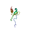
| |||||||||
|---|---|---|---|---|---|---|---|---|---|---|
| 1 |
| |||||||||
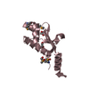
| NMR ensembles |
|
-Components
| #1: Protein | Mass: 7748.646 Da / Num. of mol.: 1 / Mutation: F13A Source method: isolated from a genetically manipulated source Source: (gene. exp.) Homo sapiens (human) / Plasmid: pET32 / Species (production host): Escherichia coli / Production host:  |
|---|
-Experimental details
-Experiment
| Experiment | Method: SOLUTION NMR | ||||||||||||||||
|---|---|---|---|---|---|---|---|---|---|---|---|---|---|---|---|---|---|
| NMR experiment |
| ||||||||||||||||
| NMR details | Text: This structure was determined using standard 3D 15N or 13C edited NMR experiments. |
- Sample preparation
| Details | Contents: 1-2mM MIP-1b F13A U-15N, 13C; 20mM Na-phosphate buffer Solvent system: 90% H2O/10% D2O |
|---|---|
| Sample conditions | Ionic strength: 20mM sodium phosphate / pH: 2.5 / Pressure: ambient / Temperature: 298 K |
| Crystal grow | *PLUS Method: other / Details: NMR |
-NMR measurement
| Radiation | Protocol: SINGLE WAVELENGTH / Monochromatic (M) / Laue (L): M | |||||||||||||||
|---|---|---|---|---|---|---|---|---|---|---|---|---|---|---|---|---|
| Radiation wavelength | Relative weight: 1 | |||||||||||||||
| NMR spectrometer |
|
- Processing
| NMR software |
| ||||||||||||||||
|---|---|---|---|---|---|---|---|---|---|---|---|---|---|---|---|---|---|
| Refinement | Method: The initial fold was obtained by distance geometry, further refined by simulated annealing. Software ordinal: 1 Details: The structure is based on a total 940 restraints, 851 distance constraints, 69 dihedral angle restraints, 20 distance restraints for 10 hydrogen bonds. | ||||||||||||||||
| NMR representative | Selection criteria: minimized average structure | ||||||||||||||||
| NMR ensemble | Conformers submitted total number: 1 |
 NMRPipe