 Movie
Movie Controller


+ Open data

- Basic information
| Entry | Database: PDB / ID: 1jdi | ||||||
|---|---|---|---|---|---|---|---|
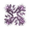
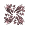
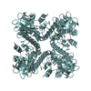
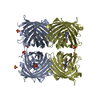
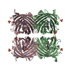
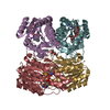
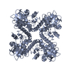
| Title | CRYSTAL STRUCTURE OF L-RIBULOSE-5-PHOSPHATE 4-EPIMERASE | ||||||
 Components Components | L-RIBULOSE 5 PHOSPHATE 4-EPIMERASE | ||||||
 Keywords Keywords | ISOMERASE / epimerase / ribulose / aldolase | ||||||
| Function / homology |  Function and homology information Function and homology informationL-ribulose-5-phosphate 4-epimerase / L-ribulose-phosphate 4-epimerase activity / L-lyxose metabolic process / : / pentose catabolic process / aldehyde-lyase activity / protein-containing complex / zinc ion binding / identical protein binding / cytosol Similarity search - Function | ||||||
| Biological species |  | ||||||
| Method |  X-RAY DIFFRACTION / SYNCHROTRON / MOLECULAR REPLACEMENT / Resolution: 2.4 Å X-RAY DIFFRACTION / SYNCHROTRON / MOLECULAR REPLACEMENT / Resolution: 2.4 Å | ||||||
 Authors Authors | Luo, Y. / Samuel, J. / Mosimann, S.C. / Lee, J.E. / Tanner, M.E. / Strynadka, N.C.J. | ||||||
 Citation Citation | Journal: Biochemistry / Year: 2001 Title: The structure of L-ribulose-5-phosphate 4-epimerase: an aldolase-like platform for epimerization. Authors: Luo, Y. / Samuel, J. / Mosimann, S.C. / Lee, J.E. / Tanner, M.E. / Strynadka, N.C. | ||||||
| History |
|
- Structure visualization
| Structure viewer | Molecule: MolmilJmol/JSmol |
|---|
- Downloads & links
-Download
| PDBx/mmCIF format | 1jdi.cif.gz | 269.1 KB | Display | PDBx/mmCIF format |
|---|---|---|---|---|
| PDB format | pdb1jdi.ent.gz | 220 KB | Display | PDB format |
| PDBx/mmJSON format | 1jdi.json.gz | Tree view | PDBx/mmJSON format | |
| Others |  Other downloads Other downloads |
-Validation report
| Arichive directory | https://data.pdbj.org/pub/pdb/validation_reports/jd/1jdiftp://data.pdbj.org/pub/pdb/validation_reports/jd/1jdi | HTTPS FTP |
|---|
-Related structure data
| Related structure data |  1k0wC  1fuaS S: Starting model for refinement C: citing same article ( |
|---|---|
| Similar structure data |


-Links
 PDBj
PDBj- Assembly
| Deposited unit | 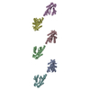
| ||||||||
|---|---|---|---|---|---|---|---|---|---|
| 1 | 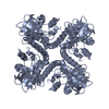
| ||||||||
| 2 | 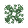
| ||||||||
| 3 | 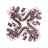
| ||||||||
| 4 | 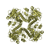
| ||||||||
| 5 | 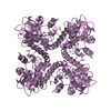
| ||||||||
| 6 | 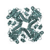
| ||||||||
| 7 | 
| ||||||||
| Unit cell |
| ||||||||
| Details | Tetramer generated by crystallographic 4-fold symmetry |
-Components
| #1: Protein | Mass: 25531.893 Da / Num. of mol.: 6 Source method: isolated from a genetically manipulated source Source: (gene. exp.) References: UniProt: P08203, L-ribulose-5-phosphate 4-epimerase #2: Chemical | ChemComp-ZN /   Mass: 65.409 Da / Num. of mol.: 6 / Source method: obtained synthetically / Formula: Zn Mass: 65.409 Da / Num. of mol.: 6 / Source method: obtained synthetically / Formula: Zn#3: Water | ChemComp-HOH / |  Mass: 18.015 Da / Num. of mol.: 433 / Source method: isolated from a natural source / Formula: H2O Mass: 18.015 Da / Num. of mol.: 433 / Source method: isolated from a natural source / Formula: H2O |
|---|
-Experimental details
-Experiment
| Experiment | Method: X-RAY DIFFRACTION / Number of used crystals: 1 |
|---|
- Sample preparation
| Crystal | Density Matthews: 2.57 Å3/Da / Density % sol: 51.7 % | ||||||||||||||||||||
|---|---|---|---|---|---|---|---|---|---|---|---|---|---|---|---|---|---|---|---|---|---|
| Crystal grow | Temperature: 295 K / Method: vapor diffusion, hanging drop / pH: 7 Details: 4.0 M sodium formate, pH 7.0, VAPOR DIFFUSION, HANGING DROP, temperature 295.0K | ||||||||||||||||||||
| Crystal grow | *PLUS | ||||||||||||||||||||
| Components of the solutions | *PLUS
|
-Data collection
| Diffraction | Mean temperature: 100 K |
|---|---|
| Diffraction source | Source: SYNCHROTRON / Site: NSLS  / Beamline: X12C / Wavelength: 0.97 Å / Beamline: X12C / Wavelength: 0.97 Å |
| Detector | Type: BRANDEIS - B4 / Detector: CCD / Date: Jul 25, 1999 |
| Radiation | Protocol: SINGLE WAVELENGTH / Monochromatic (M) / Laue (L): M / Scattering type: x-ray |
| Radiation wavelength | Wavelength: 0.97 Å / Relative weight: 1 |
| Reflection | Resolution: 2.4→30 Å / Num. all: 62146 / Num. obs: 58293 / % possible obs: 93.8 % / Observed criterion σ(F): 0 / Observed criterion σ(I): 0 / Redundancy: 3.6 % / Biso Wilson estimate: 37.8 Å2 / Rmerge(I) obs: 0.056 / Rsym value: 0.056 / Net I/σ(I): 19.4 |
| Reflection shell | Resolution: 2.4→2.49 Å / Redundancy: 3.1 % / Rmerge(I) obs: 0.201 / Mean I/σ(I) obs: 4.1 / Num. unique all: 4187 / Rsym value: 0.201 / % possible all: 68.8 |
| Reflection | *PLUS Highest resolution: 2.4 Å / Redundancy: 3.64 % |
| Reflection shell | *PLUS % possible obs: 68.8 % / Num. unique obs: 4187 |
- Processing
| Software |
| |||||||||||||||||||||||||
|---|---|---|---|---|---|---|---|---|---|---|---|---|---|---|---|---|---|---|---|---|---|---|---|---|---|---|
| Refinement | Method to determine structure: MOLECULAR REPLACEMENT Starting model: PDB ENTRY 1FUA Resolution: 2.4→15 Å / Cross valid method: THROUGHOUT / σ(F): 0 / σ(I): 0 / Stereochemistry target values: Engh & Huber
| |||||||||||||||||||||||||
| Solvent computation | Solvent model: CNS / Bsol: 37.1 Å2 / ksol: 0.399 e/Å3 | |||||||||||||||||||||||||
| Displacement parameters | Biso mean: 37.7 Å2 | |||||||||||||||||||||||||
| Refinement step | Cycle: LAST / Resolution: 2.4→15 Å
| |||||||||||||||||||||||||
| Refine LS restraints |
| |||||||||||||||||||||||||
| Software | *PLUS Name: CNS / Version: 0.9 / Classification: refinement | |||||||||||||||||||||||||
| Refinement | *PLUS Highest resolution: 2.4 Å / Num. reflection obs: 54929 / σ(F): 0 / % reflection Rfree: 10 % | |||||||||||||||||||||||||
| Solvent computation | *PLUS | |||||||||||||||||||||||||
| Displacement parameters | *PLUS Biso mean: 37.7 Å2 |
