 Movie
Movie Controller

[English] 日本語
 Yorodumi
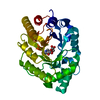
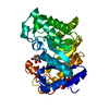
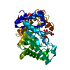
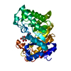
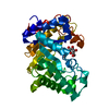
- PDB-1j01: Crystal Structure Of The Xylanase Cex With Xylobiose-Derived Inhi... -
+ Open data

- Basic information
| Entry | Database: PDB / ID: 1j01 | |||||||||
|---|---|---|---|---|---|---|---|---|---|---|
| Title | Crystal Structure Of The Xylanase Cex With Xylobiose-Derived Inhibitor Isofagomine lactam | |||||||||
 Components Components | beta-1,4-xylanase | |||||||||
 Keywords Keywords | HYDROLASE / CEX / XYLANASE / DEOXYNOJIRIMYCIN INHIBITOR / CELLULOSE DEGRADATION | |||||||||
| Function / homology |  Function and homology information Function and homology informationcellulose 1,4-beta-cellobiosidase (non-reducing end) / cellulose 1,4-beta-cellobiosidase activity / endo-1,4-beta-xylanase / endo-1,4-beta-xylanase activity / xylan catabolic process / polysaccharide binding / cellulose catabolic process Similarity search - Function | |||||||||
| Biological species |  Cellulomonas fimi (bacteria) Cellulomonas fimi (bacteria) | |||||||||
| Method |  X-RAY DIFFRACTION / MOLECULAR REPLACEMENT / Resolution: 2 Å X-RAY DIFFRACTION / MOLECULAR REPLACEMENT / Resolution: 2 Å | |||||||||
 Authors Authors | Williams, S.J. / Notenboom, V. / Wicki, J. / Rose, D.R. / Withers, S.G. | |||||||||
 Citation Citation | Journal: J.Am.Chem.Soc. / Year: 2000 Title: A New, Simple, High-Affinity Glycosidase Inhibitor: Analysis of Binding through X-ray Crystallography, Mutagenesis, and Kinetic Analysis Authors: Williams, S.J. / Notenboom, V. / Wicki, J. / Rose, D.R. / Withers, S.G. | |||||||||
| History |
|
- Structure visualization
| Structure viewer | Molecule: MolmilJmol/JSmol |
|---|
- Downloads & links
-Download
| PDBx/mmCIF format | 1j01.cif.gz | 74.9 KB | Display | PDBx/mmCIF format |
|---|---|---|---|---|
| PDB format | pdb1j01.ent.gz | 55.7 KB | Display | PDB format |
| PDBx/mmJSON format | 1j01.json.gz | Tree view | PDBx/mmJSON format | |
| Others |  Other downloads Other downloads |
-Validation report
| Arichive directory | https://data.pdbj.org/pub/pdb/validation_reports/j0/1j01ftp://data.pdbj.org/pub/pdb/validation_reports/j0/1j01 | HTTPS FTP |
|---|
-Related structure data
| Related structure data | |
|---|---|
| Similar structure data |


-Links
 PDBj
PDBj

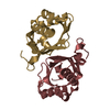
- Assembly
| Deposited unit | 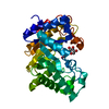
| ||||||||
|---|---|---|---|---|---|---|---|---|---|
| 1 |
| ||||||||
| Unit cell |
|
-Components
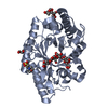
| #1: Protein | Mass: 34051.941 Da / Num. of mol.: 1 / Fragment: catalytic domain Source method: isolated from a genetically manipulated source Source: (gene. exp.) Cellulomonas fimi (bacteria) / Gene: Cex / Plasmid: pUC12 / Production host: References: UniProt: P07986, cellulose 1,4-beta-cellobiosidase (non-reducing end) |
|---|---|
| #2: Sugar | ChemComp-XIL / (  Type: D-saccharide / Mass: 263.244 Da / Num. of mol.: 1 / Source method: obtained synthetically / Formula: C10H17NO7 Type: D-saccharide / Mass: 263.244 Da / Num. of mol.: 1 / Source method: obtained synthetically / Formula: C10H17NO7 |
| #3: Water | ChemComp-HOH /  Mass: 18.015 Da / Num. of mol.: 160 / Source method: isolated from a natural source / Formula: H2O Mass: 18.015 Da / Num. of mol.: 160 / Source method: isolated from a natural source / Formula: H2O |
| Has protein modification | Y |
-Experimental details
-Experiment
| Experiment | Method: X-RAY DIFFRACTION / Number of used crystals: 1 |
|---|
- Sample preparation
| Crystal | Density Matthews: 2.23 Å3/Da / Density % sol: 44.89 % | |||||||||||||||||||||
|---|---|---|---|---|---|---|---|---|---|---|---|---|---|---|---|---|---|---|---|---|---|---|
| Crystal grow | Temperature: 298 K / Method: vapor diffusion, hanging drop / pH: 4.6 Details: PEG4000, sodium acetate, pH 4.6, VAPOR DIFFUSION, HANGING DROP, temperature 298K | |||||||||||||||||||||
| Crystal grow | *PLUS Method: unknown | |||||||||||||||||||||
| Components of the solutions | *PLUS
|
-Data collection
| Diffraction | Mean temperature: 100 K |
|---|---|
| Diffraction source | Source: ROTATING ANODE / Type: RIGAKU RU200 / Wavelength: 1.54 Å |
| Detector | Type: MARRESEARCH / Detector: IMAGE PLATE / Date: Oct 1, 2000 |
| Radiation | Monochromator: Osmic focussing optics / Protocol: SINGLE WAVELENGTH / Monochromatic (M) / Laue (L): M / Scattering type: x-ray |
| Radiation wavelength | Wavelength: 1.54 Å / Relative weight: 1 |
| Reflection | Resolution: 2→50 Å / Num. all: 21434 / Num. obs: 20862 / % possible obs: 97.3 % / Observed criterion σ(F): 0 / Observed criterion σ(I): 0 |
| Reflection shell | Resolution: 2→2.1 Å / % possible all: 85 |
| Reflection | *PLUS Num. obs: 21259 / % possible obs: 99 % / Num. measured all: 258208 / Rmerge(I) obs: 0.057 |
- Processing
| Software |
| ||||||||||||||||||||
|---|---|---|---|---|---|---|---|---|---|---|---|---|---|---|---|---|---|---|---|---|---|
| Refinement | Method to determine structure: MOLECULAR REPLACEMENT / Resolution: 2→500 Å / σ(F): 0 / Stereochemistry target values: Engh & Huber
| ||||||||||||||||||||
| Refinement step | Cycle: LAST / Resolution: 2→500 Å
| ||||||||||||||||||||
| Refinement | *PLUS Lowest resolution: 500 Å / Rfactor Rfree: 0.25 | ||||||||||||||||||||
| Solvent computation | *PLUS | ||||||||||||||||||||
| Displacement parameters | *PLUS |
