 Movie
Movie Controller

[English] 日本語
 Yorodumi
- PDB-1iw8: Crystal Structure of a mutant of acid phosphatase from Escherichi... -
+ Open data

- Basic information
| Entry | Database: PDB / ID: 1iw8 | ||||||
|---|---|---|---|---|---|---|---|
| Title | Crystal Structure of a mutant of acid phosphatase from Escherichia blattae (G74D/I153T) | ||||||
 Components Components | acid phosphatase | ||||||
 Keywords Keywords | HYDROLASE / ALL ALPHA | ||||||
| Function / homology |  Function and homology information Function and homology informationacid phosphatase / acid phosphatase activity / outer membrane-bounded periplasmic space Similarity search - Function | ||||||
| Biological species |  Escherichia blattae (bacteria) Escherichia blattae (bacteria) | ||||||
| Method |  X-RAY DIFFRACTION / SYNCHROTRON / MOLECULAR REPLACEMENT / Resolution: 2.5 Å X-RAY DIFFRACTION / SYNCHROTRON / MOLECULAR REPLACEMENT / Resolution: 2.5 Å | ||||||
 Authors Authors | Ishikawa, K. / Mihara, Y. / Shimba, N. / Ohtsu, N. / Kawasaki, H. / Suzuki, E. / Asano, Y. | ||||||
 Citation Citation | Journal: PROTEIN ENG. / Year: 2002 Title: Enhancement of nucleoside phosphorylation activity in an acid phosphatase Authors: Ishikawa, K. / Mihara, Y. / Shimba, N. / Ohtsu, N. / Kawasaki, H. / Suzuki, E. / Asano, Y. #1: Journal: Embo J. / Year: 2000Title: X-ray structures of a novel acid phosphatase from Escherichia blattae and its complex with the transition-state analog molybdate Authors: Ishikawa, K. / Mihara, Y. / Gondoh, K. / Suzuki, E. / Asano, Y. | ||||||
| History |
|
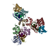
- Structure visualization
| Structure viewer | Molecule: MolmilJmol/JSmol |
|---|
- Downloads & links
-Download
| PDBx/mmCIF format | 1iw8.cif.gz | 253.8 KB | Display | PDBx/mmCIF format |
|---|---|---|---|---|
| PDB format | pdb1iw8.ent.gz | 207.4 KB | Display | PDB format |
| PDBx/mmJSON format | 1iw8.json.gz | Tree view | PDBx/mmJSON format | |
| Others |  Other downloads Other downloads |
-Validation report
| Arichive directory | https://data.pdbj.org/pub/pdb/validation_reports/iw/1iw8ftp://data.pdbj.org/pub/pdb/validation_reports/iw/1iw8 | HTTPS FTP |
|---|
-Related structure data
| Related structure data |  1d2tS S: Starting model for refinement |
|---|---|
| Similar structure data |


-Links
 PDBj
PDBj
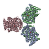
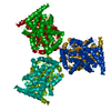
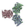
- Assembly
| Deposited unit | 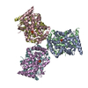
| ||||||||
|---|---|---|---|---|---|---|---|---|---|
| 1 |
| ||||||||
| Unit cell |
|
-Components
| #1: Protein | Mass: 25079.078 Da / Num. of mol.: 6 / Fragment: residues 1-231 / Mutation: G74D, I153T Source method: isolated from a genetically manipulated source Source: (gene. exp.) Escherichia blattae (bacteria) / Plasmid: pUC118 / Production host: #2: Chemical | ChemComp-SO4 /   Mass: 96.063 Da / Num. of mol.: 6 / Source method: obtained synthetically / Formula: SO4 Mass: 96.063 Da / Num. of mol.: 6 / Source method: obtained synthetically / Formula: SO4#3: Water | ChemComp-HOH / |  Mass: 18.015 Da / Num. of mol.: 314 / Source method: isolated from a natural source / Formula: H2O Mass: 18.015 Da / Num. of mol.: 314 / Source method: isolated from a natural source / Formula: H2OHas protein modification | Y | |
|---|
-Experimental details
-Experiment
| Experiment | Method: X-RAY DIFFRACTION / Number of used crystals: 2 |
|---|
- Sample preparation
| Crystal | Density Matthews: 2.25 Å3/Da / Density % sol: 45.28 % | ||||||||||||||||||||||||||||||
|---|---|---|---|---|---|---|---|---|---|---|---|---|---|---|---|---|---|---|---|---|---|---|---|---|---|---|---|---|---|---|---|
| Crystal grow | Temperature: 293 K / Method: vapor diffusion, hanging drop / pH: 8.4 Details: PEG400, pH 8.4, VAPOR DIFFUSION, HANGING DROP, temperature 293K | ||||||||||||||||||||||||||||||
| Crystal grow | *PLUS Temperature: 20 ℃ / pH: 8 | ||||||||||||||||||||||||||||||
| Components of the solutions | *PLUS
|
-Data collection
| Diffraction | Mean temperature: 288 K |
|---|---|
| Diffraction source | Source: SYNCHROTRON / Site: Photon Factory  / Beamline: BL-6B / Wavelength: 1 Å / Beamline: BL-6B / Wavelength: 1 Å |
| Detector | Type: FUJI / Detector: IMAGE PLATE / Date: Dec 3, 1997 |
| Radiation | Protocol: SINGLE WAVELENGTH / Monochromatic (M) / Laue (L): M / Scattering type: x-ray |
| Radiation wavelength | Wavelength: 1 Å / Relative weight: 1 |
| Reflection | Resolution: 2.5→10 Å / Num. all: 45889 / Num. obs: 45889 / % possible obs: 97.5 % / Observed criterion σ(F): 1 / Observed criterion σ(I): 1 / Biso Wilson estimate: 28.6 Å2 / Rmerge(I) obs: 0.084 |
| Reflection shell | Resolution: 2.5→2.55 Å / % possible all: 91.2 |
| Reflection | *PLUS Lowest resolution: 10 Å |
- Processing
| Software |
| |||||||||||||||||||||||||
|---|---|---|---|---|---|---|---|---|---|---|---|---|---|---|---|---|---|---|---|---|---|---|---|---|---|---|
| Refinement | Method to determine structure: MOLECULAR REPLACEMENT Starting model: PDB ENTRY 1D2T Resolution: 2.5→10 Å / Isotropic thermal model: Isotropic / σ(F): 1 / Stereochemistry target values: Engh & Huber
| |||||||||||||||||||||||||
| Displacement parameters | Biso mean: 36.3 Å2 | |||||||||||||||||||||||||
| Refine analyze | Luzzati coordinate error obs: 0.32 Å / Luzzati d res low obs: 5 Å / Luzzati sigma a obs: 0.48 Å | |||||||||||||||||||||||||
| Refinement step | Cycle: LAST / Resolution: 2.5→10 Å
| |||||||||||||||||||||||||
| Refine LS restraints |
| |||||||||||||||||||||||||
| LS refinement shell | Resolution: 2.5→2.65 Å / Rfactor Rfree error: 0.021
| |||||||||||||||||||||||||
| Refinement | *PLUS Lowest resolution: 10 Å / σ(F): 1 / Rfactor Rfree: 0.285 / Rfactor Rwork: 0.242 | |||||||||||||||||||||||||
| Solvent computation | *PLUS | |||||||||||||||||||||||||
| Displacement parameters | *PLUS | |||||||||||||||||||||||||
| Refine LS restraints | *PLUS Type: c_angle_deg / Dev ideal: 1.31 |
