 Movie
Movie Controller

[English] 日本語
 Yorodumi
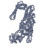
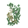
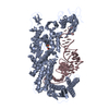
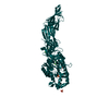
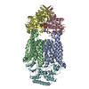
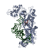
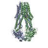
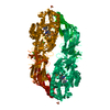
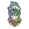
- PDB-1ivs: CRYSTAL STRUCTURE OF THERMUS THERMOPHILUS VALYL-TRNA SYNTHETASE C... -
+ Open data

- Basic information
| Entry | Database: PDB / ID: 1ivs | ||||||
|---|---|---|---|---|---|---|---|
| Title | CRYSTAL STRUCTURE OF THERMUS THERMOPHILUS VALYL-TRNA SYNTHETASE COMPLEXED WITH TRNA(VAL) AND VALYL-ADENYLATE ANALOGUE | ||||||
 Components Components |
| ||||||
 Keywords Keywords | LIGASE/RNA / Rossmann fold / helix bundle / coiled coil / beta barrel / RIKEN Structural Genomics/Proteomics Initiative / RSGI / Structural Genomics / LIGASE-RNA COMPLEX | ||||||
| Function / homology |  Function and homology information Function and homology informationvaline-tRNA ligase / valine-tRNA ligase activity / valyl-tRNA aminoacylation / aminoacyl-tRNA deacylase activity / ATP binding / metal ion binding / cytosol Similarity search - Function | ||||||
| Biological species |   Thermus thermophilus (bacteria) Thermus thermophilus (bacteria) | ||||||
| Method |  X-RAY DIFFRACTION / SYNCHROTRON / MIR / Resolution: 2.9 Å X-RAY DIFFRACTION / SYNCHROTRON / MIR / Resolution: 2.9 Å | ||||||
 Authors Authors | Fukai, S. / Nureki, O. / Sekine, S.-I. / Shimada, A. / Vassylyev, D.G. / Yokoyama, S. / RIKEN Structural Genomics/Proteomics Initiative (RSGI) | ||||||
 Citation Citation | Journal: RNA / Year: 2003 Title: Mechanism of molecular interactions for tRNA(Val) recognition by valyl-tRNA synthetase Authors: Fukai, S. / Nureki, O. / Sekine, S.-I. / Shimada, A. / Vassylyev, D.G. / Yokoyama, S. #1: Journal: Cell(Cambridge,Mass.) / Year: 2000Title: STRUCTURAL BASIS FOR DOUBLE-SIEVE DISCRIMINATION OF L-VALINE FROM L-ISOLEUCINE AND L-THREONINE BY THE COMPLEX OF TRNA(VAL) AND VALYL-TRNA SYNTHETASE Authors: Fukai, S. / Nureki, O. / Sekine, S. / Shimada, A. / Tao, J. / Vassylyev, D.G. / Yokoyama, S. | ||||||
| History |
|
- Structure visualization
| Structure viewer | Molecule: MolmilJmol/JSmol |
|---|
- Downloads & links
-Download
| PDBx/mmCIF format | 1ivs.cif.gz | 441.4 KB | Display | PDBx/mmCIF format |
|---|---|---|---|---|
| PDB format | pdb1ivs.ent.gz | 352.3 KB | Display | PDB format |
| PDBx/mmJSON format | 1ivs.json.gz | Tree view | PDBx/mmJSON format | |
| Others |  Other downloads Other downloads |
-Validation report
| Arichive directory | https://data.pdbj.org/pub/pdb/validation_reports/iv/1ivsftp://data.pdbj.org/pub/pdb/validation_reports/iv/1ivs | HTTPS FTP |
|---|
-Related structure data


-Links
 PDBj
PDBj


- Assembly
| Deposited unit | 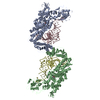
| ||||||||
|---|---|---|---|---|---|---|---|---|---|
| 1 | 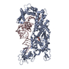
| ||||||||
| 2 | 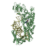
| ||||||||
| Unit cell |
|
-Components
| #1: RNA chain | Mass: 24174.455 Da / Num. of mol.: 2 / Source method: obtained synthetically / Details: tRNA (Val) with the CAC anticodon #2: Protein | Mass: 98914.445 Da / Num. of mol.: 2 Source method: isolated from a genetically manipulated source Source: (gene. exp.) Thermus thermophilus (bacteria) / Gene: valS / Plasmid: pK7 / Production host: #3: Chemical | 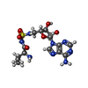  Mass: 444.466 Da / Num. of mol.: 2 / Source method: obtained synthetically / Formula: C15H24N8O6S Mass: 444.466 Da / Num. of mol.: 2 / Source method: obtained synthetically / Formula: C15H24N8O6S#4: Water | ChemComp-HOH / |  Mass: 18.015 Da / Num. of mol.: 218 / Source method: isolated from a natural source / Formula: H2O Mass: 18.015 Da / Num. of mol.: 218 / Source method: isolated from a natural source / Formula: H2O |
|---|
-Experimental details
-Experiment
| Experiment | Method: X-RAY DIFFRACTION / Number of used crystals: 1 |
|---|
- Sample preparation
| Crystal grow | Temperature: 277 K / Method: vapor diffusion, hanging drop / pH: 6.5 Details: Ammonium Sulfate, Magnesium Sulfate, Cacodylate Na, 1,8-diaminooctane, pH 6.5, VAPOR DIFFUSION, HANGING DROP, temperature 277K | |||||||||||||||||||||||||||||||||||||||||||||
|---|---|---|---|---|---|---|---|---|---|---|---|---|---|---|---|---|---|---|---|---|---|---|---|---|---|---|---|---|---|---|---|---|---|---|---|---|---|---|---|---|---|---|---|---|---|---|
| Crystal grow | *PLUS Temperature: 4 ℃Details: Fukai, S., (2000) Cell (Cambridge,Mass.), 103, 793. | |||||||||||||||||||||||||||||||||||||||||||||
| Components of the solutions | *PLUS
|
-Data collection
| Diffraction | Mean temperature: 90 K |
|---|---|
| Diffraction source | Source: SYNCHROTRON / Site: SPring-8  / Beamline: BL41XU / Wavelength: 0.708 Å / Beamline: BL41XU / Wavelength: 0.708 Å |
| Detector | Type: RIGAKU RAXIS IV / Detector: IMAGE PLATE / Date: Apr 24, 1998 |
| Radiation | Monochromator: Si(111) / Protocol: SINGLE WAVELENGTH / Monochromatic (M) / Laue (L): M / Scattering type: x-ray |
| Radiation wavelength | Wavelength: 0.708 Å / Relative weight: 1 |
| Reflection | Resolution: 2.9→50 Å / Num. all: 150310 / Num. obs: 150310 / % possible obs: 96.5 % / Observed criterion σ(F): 0 / Observed criterion σ(I): 0 / Biso Wilson estimate: 56.5 Å2 |
| Reflection shell | Resolution: 2.9→3.08 Å / % possible all: 87.9 |
| Reflection | *PLUS |
- Processing
| Software |
| ||||||||||||||||||||||||||||||||||||
|---|---|---|---|---|---|---|---|---|---|---|---|---|---|---|---|---|---|---|---|---|---|---|---|---|---|---|---|---|---|---|---|---|---|---|---|---|---|
| Refinement | Method to determine structure: MIR / Resolution: 2.9→40 Å / Rfactor Rfree error: 0.003 / Isotropic thermal model: RESTRAINED / Cross valid method: THROUGHOUT / σ(F): 0 / Stereochemistry target values: Engh & Huber
| ||||||||||||||||||||||||||||||||||||
| Solvent computation | Solvent model: FLAT MODEL / Bsol: 25.6469 Å2 / ksol: 0.297208 e/Å3 | ||||||||||||||||||||||||||||||||||||
| Displacement parameters | Biso mean: 50.8 Å2
| ||||||||||||||||||||||||||||||||||||
| Refine analyze |
| ||||||||||||||||||||||||||||||||||||
| Refinement step | Cycle: LAST / Resolution: 2.9→40 Å
| ||||||||||||||||||||||||||||||||||||
| Refine LS restraints |
| ||||||||||||||||||||||||||||||||||||
| LS refinement shell | Resolution: 2.9→3.08 Å / Rfactor Rfree error: 0.011 / Total num. of bins used: 6
| ||||||||||||||||||||||||||||||||||||
| Xplor file |
| ||||||||||||||||||||||||||||||||||||
| Refinement | *PLUS Highest resolution: 2.9 Å / % reflection Rfree: 5 % / Rfactor Rfree: 0.281 / Rfactor Rwork: 0.247 | ||||||||||||||||||||||||||||||||||||
| Solvent computation | *PLUS | ||||||||||||||||||||||||||||||||||||
| Displacement parameters | *PLUS | ||||||||||||||||||||||||||||||||||||
| Refine LS restraints | *PLUS
|
