- PDB-1ian: HUMAN P38 MAP KINASE INHIBITOR COMPLEX -
+
Open data
ID or keywords:
Loading...
-
Basic information
Entry
Database: PDB / ID: 1ian
Title
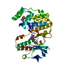
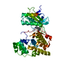
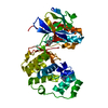
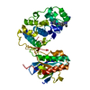
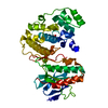
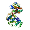
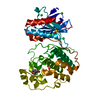
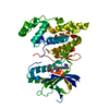
HUMAN P38 MAP KINASE INHIBITOR COMPLEX
Components
P38 MAP KINASE
Keywords
SERINE/THREONINE-PROTEIN KINASE / PROTEIN SER/THR-KINASE / SERINE-THREONINE-PROTEIN KINASE complex
Function / homology
Function and homology information
positive regulation of cyclase activity / Activation of PPARGC1A (PGC-1alpha) by phosphorylation / regulation of synaptic membrane adhesion / stress-induced premature senescence / stress-activated protein kinase signaling cascade / CD163 mediating an anti-inflammatory response / 3'-UTR-mediated mRNA stabilization / cell surface receptor protein serine/threonine kinase signaling pathway / positive regulation of myoblast fusion / KSRP (KHSRP) binds and destabilizes mRNA ...positive regulation of cyclase activity / Activation of PPARGC1A (PGC-1alpha) by phosphorylation / regulation of synaptic membrane adhesion / stress-induced premature senescence / stress-activated protein kinase signaling cascade / CD163 mediating an anti-inflammatory response / 3'-UTR-mediated mRNA stabilization / cell surface receptor protein serine/threonine kinase signaling pathway / positive regulation of myoblast fusion / KSRP (KHSRP) binds and destabilizes mRNA / : / cellular response to UV-B / cartilage condensation / mitogen-activated protein kinase p38 binding / positive regulation of muscle cell differentiation / Myogenesis / Platelet sensitization by LDL / NFAT protein binding / positive regulation of myotube differentiation / regulation of cytokine production involved in inflammatory response / Activation of the AP-1 family of transcription factors / ERK/MAPK targets / cellular response to lipoteichoic acid / p38MAPK cascade / response to dietary excess / fatty acid oxidation / response to muramyl dipeptide / MAP kinase kinase activity / Regulation of MITF-M-dependent genes involved in pigmentation / MAP kinase activity / cellular response to vascular endothelial growth factor stimulus / regulation of ossification / mitogen-activated protein kinase / vascular endothelial growth factor receptor signaling pathway / chondrocyte differentiation / RHO GTPases Activate NADPH Oxidases / positive regulation of myoblast differentiation / negative regulation of hippo signaling / stress-activated MAPK cascade / positive regulation of cardiac muscle cell proliferation / skeletal muscle tissue development / positive regulation of brown fat cell differentiation / response to muscle stretch / p38MAPK events / signal transduction in response to DNA damage / striated muscle cell differentiation / positive regulation of interleukin-12 production / positive regulation of erythrocyte differentiation / osteoclast differentiation / lipopolysaccharide-mediated signaling pathway / DNA damage checkpoint signaling / placenta development / tumor necrosis factor-mediated signaling pathway / positive regulation of D-glucose import across plasma membrane / stem cell differentiation / cellular response to ionizing radiation / activated TAK1 mediates p38 MAPK activation / negative regulation of inflammatory response to antigenic stimulus / negative regulation of canonical Wnt signaling pathway / NOD1/2 Signaling Pathway / platelet activation / cellular response to virus / positive regulation of protein import into nucleus / response to insulin / bone development / VEGFA-VEGFR2 Pathway / glucose metabolic process / positive regulation of reactive oxygen species metabolic process / cell morphogenesis / chemotaxis / spindle pole / cellular senescence / osteoblast differentiation / ADP signalling through P2Y purinoceptor 1 / MAPK cascade / cellular response to lipopolysaccharide / angiogenesis / secretory granule lumen / protein phosphatase binding / Oxidative Stress Induced Senescence / transcription by RNA polymerase II / Regulation of TP53 Activity through Phosphorylation / ficolin-1-rich granule lumen / cell surface receptor signaling pathway / nuclear speck / intracellular signal transduction / protein serine kinase activity / protein serine/threonine kinase activity / apoptotic process / Neutrophil degranulation / positive regulation of gene expression / regulation of transcription by RNA polymerase II / glutamatergic synapse / enzyme binding / signal transduction / positive regulation of transcription by RNA polymerase II / mitochondrion / extracellular region / nucleoplasm / ATP binding Similarity search - Function
Mitogen-activated protein kinase 14 / Mitogen-activated protein (MAP) kinase p38-like / Mitogen-activated protein (MAP) kinase, conserved site / MAP kinase signature. / : / Protein kinase domain / Serine/Threonine protein kinases, catalytic domain / Protein kinase, ATP binding site / Protein kinases ATP-binding region signature. / Protein kinase domain profile. ...Mitogen-activated protein kinase 14 / Mitogen-activated protein (MAP) kinase p38-like / Mitogen-activated protein (MAP) kinase, conserved site / MAP kinase signature. / : / Protein kinase domain / Serine/Threonine protein kinases, catalytic domain / Protein kinase, ATP binding site / Protein kinases ATP-binding region signature. / Protein kinase domain profile. / Protein kinase domain / Protein kinase-like domain superfamily Similarity search - Domain/homology
In the structure databanks used in Yorodumi, some data are registered as the other names, "COVID-19 virus" and "2019-nCoV". Here are the details of the virus and the list of structure data.
Jan 31, 2019. EMDB accession codes are about to change! (news from PDBe EMDB page)
EMDB accession codes are about to change! (news from PDBe EMDB page)
The allocation of 4 digits for EMDB accession codes will soon come to an end. Whilst these codes will remain in use, new EMDB accession codes will include an additional digit and will expand incrementally as the available range of codes is exhausted. The current 4-digit format prefixed with “EMD-” (i.e. EMD-XXXX) will advance to a 5-digit format (i.e. EMD-XXXXX), and so on. It is currently estimated that the 4-digit codes will be depleted around Spring 2019, at which point the 5-digit format will come into force.
The EM Navigator/Yorodumi systems omit the EMD- prefix.
Related info.:Q: What is EMD? / ID/Accession-code notation in Yorodumi/EM Navigator
Yorodumi is a browser for structure data from EMDB, PDB, SASBDB, etc.
This page is also the successor to EM Navigator detail page, and also detail information page/front-end page for Omokage search.
The word "yorodu" (or yorozu) is an old Japanese word meaning "ten thousand". "mi" (miru) is to see.
Related info.:EMDB / PDB / SASBDB / Comparison of 3 databanks / Yorodumi Search / Aug 31, 2016. New EM Navigator & Yorodumi / Yorodumi Papers / Jmol/JSmol / Function and homology information / Changes in new EM Navigator and Yorodumi
 Movie
Movie Controller


 Open data
 Basic information
 Components
 Keywords
 Function and homology information
 Homo sapiens (human)
 X-RAY DIFFRACTION /
  Authors
 Citation
 Structure visualization
 Downloads & links
 Other downloads
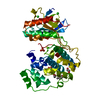

 PDBj
PDBj


 Assembly
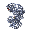


 Mass: 485.341 Da / Num. of mol.: 3 / Source method: obtained synthetically / Formula: C21H16IN3OS
 Sample preparation
 / Beamline: X4A / Wavelength: 0.9792
  Processing