 Movie
Movie Controller

[English] 日本語
 Yorodumi
- PDB-1i75: CRYSTAL STRUCTURE OF CYCLODEXTRIN GLUCANOTRANSFERASE FROM ALKALOP... -
+ Open data

- Basic information
| Entry | Database: PDB / ID: 1i75 | ||||||
|---|---|---|---|---|---|---|---|
| Title | CRYSTAL STRUCTURE OF CYCLODEXTRIN GLUCANOTRANSFERASE FROM ALKALOPHILIC BACILLUS SP.#1011 COMPLEXED WITH 1-DEOXYNOJIRIMYCIN | ||||||
 Components Components | CYCLODEXTRIN GLUCANOTRANSFERASE | ||||||
 Keywords Keywords | TRANSFERASE / PROTEIN-INHIBITOR COMPLEX | ||||||
| Function / homology |  Function and homology information Function and homology informationcyclomaltodextrin glucanotransferase / cyclomaltodextrin glucanotransferase activity / starch binding / alpha-amylase activity / carbohydrate metabolic process / extracellular region / metal ion binding Similarity search - Function | ||||||
| Biological species |  | ||||||
| Method |  X-RAY DIFFRACTION / MOLECULAR REPLACEMENT / Resolution: 2 Å X-RAY DIFFRACTION / MOLECULAR REPLACEMENT / Resolution: 2 Å | ||||||
 Authors Authors | Kanai, R. / Haga, K. / Yamane, K. / Harata, K. | ||||||
 Citation Citation | Journal: J.Biochem.(Tokyo) / Year: 2001 Title: Crystal structure of cyclodextrin glucanotransferase from alkalophilic Bacillus sp. 1011 complexed with 1-deoxynojirimycin at 2.0 A resolution. Authors: Kanai, R. / Haga, K. / Yamane, K. / Harata, K. #1: Journal: Acta Crystallogr.,Sect.D / Year: 1996Title: X-ray Structure of Cyclodextrin Glucanotransferase from Alkalophilic Bacillus Sp.1011. Comparison of Two Independent Molecules at 1.8 A Resolution | ||||||
| History |
|
- Structure visualization
| Structure viewer | Molecule: MolmilJmol/JSmol |
|---|
- Downloads & links
-Download
| PDBx/mmCIF format | 1i75.cif.gz | 285.5 KB | Display | PDBx/mmCIF format |
|---|---|---|---|---|
| PDB format | pdb1i75.ent.gz | 227.6 KB | Display | PDB format |
| PDBx/mmJSON format | 1i75.json.gz | Tree view | PDBx/mmJSON format | |
| Others |  Other downloads Other downloads |
-Validation report
| Arichive directory | https://data.pdbj.org/pub/pdb/validation_reports/i7/1i75ftp://data.pdbj.org/pub/pdb/validation_reports/i7/1i75 | HTTPS FTP |
|---|
-Related structure data
| Related structure data |  1pamS S: Starting model for refinement |
|---|---|
| Similar structure data |


-Links
 PDBj
PDBj


- Assembly
| Deposited unit | 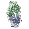
| ||||||||
|---|---|---|---|---|---|---|---|---|---|
| 1 |
| ||||||||
| Unit cell |
|
-Components
| #1: Protein | Mass: 75230.086 Da / Num. of mol.: 2 Source method: isolated from a genetically manipulated source Source: (gene. exp.) References: UniProt: P05618, cyclomaltodextrin glucanotransferase #2: Chemical | ChemComp-CA /   Mass: 40.078 Da / Num. of mol.: 4 / Source method: obtained synthetically / Formula: Ca Mass: 40.078 Da / Num. of mol.: 4 / Source method: obtained synthetically / Formula: Ca#3: Sugar | ChemComp-NOJ /   Type: D-saccharide / Mass: 163.172 Da / Num. of mol.: 4 / Source method: obtained synthetically / Formula: C6H13NO4 Type: D-saccharide / Mass: 163.172 Da / Num. of mol.: 4 / Source method: obtained synthetically / Formula: C6H13NO4#4: Water | ChemComp-HOH / |  Mass: 18.015 Da / Num. of mol.: 590 / Source method: isolated from a natural source / Formula: H2O Mass: 18.015 Da / Num. of mol.: 590 / Source method: isolated from a natural source / Formula: H2OHas protein modification | Y | |
|---|
-Experimental details
-Experiment
| Experiment | Method: X-RAY DIFFRACTION / Number of used crystals: 2 |
|---|
- Sample preparation
| Crystal | Density Matthews: 2.39 Å3/Da / Density % sol: 48.61 % | |||||||||||||||||||||||||||||||||||
|---|---|---|---|---|---|---|---|---|---|---|---|---|---|---|---|---|---|---|---|---|---|---|---|---|---|---|---|---|---|---|---|---|---|---|---|---|
| Crystal grow | Temperature: 298 K / Method: vapor diffusion, hanging drop / pH: 5.6 Details: PEG3000, 2-Propanol, sodium citrate, calcium chloride, 1-deoxynojirimycin, pH 5.6, VAPOR DIFFUSION, HANGING DROP, temperature 298K | |||||||||||||||||||||||||||||||||||
| Crystal grow | *PLUS | |||||||||||||||||||||||||||||||||||
| Components of the solutions | *PLUS
|
-Data collection
| Diffraction | Mean temperature: 286 K |
|---|---|
| Diffraction source | Source: ROTATING ANODE / Type: ENRAF-NONIUS FR571 / Wavelength: 1.5418 Å |
| Detector | Type: ENRAF-NONIUS FAST / Detector: DIFFRACTOMETER / Date: Dec 20, 1997 / Details: graphite monochromator |
| Radiation | Monochromator: GRAPHITE / Protocol: SINGLE WAVELENGTH / Monochromatic (M) / Laue (L): M / Scattering type: x-ray |
| Radiation wavelength | Wavelength: 1.5418 Å / Relative weight: 1 |
| Reflection | Resolution: 1.84→27.89 Å / Num. all: 106410 / Num. obs: 106410 / % possible obs: 81.7 % / Observed criterion σ(F): 0 / Observed criterion σ(I): 0 / Redundancy: 3 % / Rmerge(I) obs: 0.076 |
| Reflection shell | Resolution: 1.84→1.87 Å / Rmerge(I) obs: 0.206 |
- Processing
| Software |
| ||||||||||||||||||||||||
|---|---|---|---|---|---|---|---|---|---|---|---|---|---|---|---|---|---|---|---|---|---|---|---|---|---|
| Refinement | Method to determine structure: MOLECULAR REPLACEMENT Starting model: PDB ENTRY 1PAM Resolution: 2→10 Å / Isotropic thermal model: Isotropic / σ(F): 2 / Stereochemistry target values: CHARMM
| ||||||||||||||||||||||||
| Refine analyze | Luzzati coordinate error obs: 0.2 Å | ||||||||||||||||||||||||
| Refinement step | Cycle: LAST / Resolution: 2→10 Å
| ||||||||||||||||||||||||
| Refine LS restraints |
| ||||||||||||||||||||||||
| LS refinement shell | Resolution: 2→2.03 Å
|
