 Movie
Movie Controller

[English] 日本語
 Yorodumi
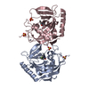
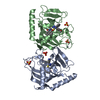
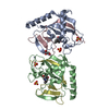
- PDB-1i5e: CRYSTAL STRUCTURE OF BACILLUS CALDOLYTICUS URACIL PHOSPHORIBOSYLT... -
+ Open data

- Basic information
| Entry | Database: PDB / ID: 1i5e | ||||||
|---|---|---|---|---|---|---|---|
| Title | CRYSTAL STRUCTURE OF BACILLUS CALDOLYTICUS URACIL PHOSPHORIBOSYLTRANSFERASE WITH BOUND UMP | ||||||
 Components Components | URACIL PHOSPHORIBOSYLTRANSFERASE | ||||||
 Keywords Keywords | TRANSFERASE / uracil phosphoribosyltransferase / salvage pathway / bacillus caldolyticus | ||||||
| Function / homology |  Function and homology information Function and homology informationuracil salvage / uracil phosphoribosyltransferase / uracil phosphoribosyltransferase activity / UMP salvage / GTP binding / magnesium ion binding / cytoplasm Similarity search - Function | ||||||
| Biological species |  Bacillus caldolyticus (bacteria) Bacillus caldolyticus (bacteria) | ||||||
| Method |  X-RAY DIFFRACTION / SYNCHROTRON / MIR / Resolution: 3 Å X-RAY DIFFRACTION / SYNCHROTRON / MIR / Resolution: 3 Å | ||||||
 Authors Authors | Kadziola, A. / Neuhard, J. / Larsen, S. | ||||||
 Citation Citation | Journal: Acta Crystallogr.,Sect.D / Year: 2002 Title: Structure of product-bound Bacillus caldolyticus uracil phosphoribosyltransferase confirms ordered sequential substrate binding. Authors: Kadziola, A. / Neuhard, J. / Larsen, S. | ||||||
| History |
|
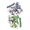
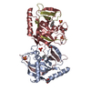
- Structure visualization
| Structure viewer | Molecule: MolmilJmol/JSmol |
|---|
- Downloads & links
-Download
| PDBx/mmCIF format | 1i5e.cif.gz | 90.1 KB | Display | PDBx/mmCIF format |
|---|---|---|---|---|
| PDB format | pdb1i5e.ent.gz | 69.2 KB | Display | PDB format |
| PDBx/mmJSON format | 1i5e.json.gz | Tree view | PDBx/mmJSON format | |
| Others |  Other downloads Other downloads |
-Validation report
| Arichive directory | https://data.pdbj.org/pub/pdb/validation_reports/i5/1i5eftp://data.pdbj.org/pub/pdb/validation_reports/i5/1i5e | HTTPS FTP |
|---|
-Related structure data
| Related structure data | |
|---|---|
| Similar structure data |


-Links
 PDBj
PDBj


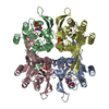
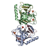
- Assembly
| Deposited unit | 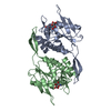
| ||||||||
|---|---|---|---|---|---|---|---|---|---|
| 1 |
| ||||||||
| Unit cell |
|
-Components
| #1: Protein | Mass: 22864.732 Da / Num. of mol.: 2 Source method: isolated from a genetically manipulated source Source: (gene. exp.) Bacillus caldolyticus (bacteria) / Gene: UPP / Production host: References: UniProt: P70881, uracil phosphoribosyltransferase #2: Chemical |   Mass: 324.181 Da / Num. of mol.: 2 / Source method: obtained synthetically / Formula: C9H13N2O9P Mass: 324.181 Da / Num. of mol.: 2 / Source method: obtained synthetically / Formula: C9H13N2O9P#3: Water | ChemComp-HOH / |  Mass: 18.015 Da / Num. of mol.: 2 / Source method: isolated from a natural source / Formula: H2O Mass: 18.015 Da / Num. of mol.: 2 / Source method: isolated from a natural source / Formula: H2O |
|---|
-Experimental details
-Experiment
| Experiment | Method: X-RAY DIFFRACTION / Number of used crystals: 1 |
|---|
- Sample preparation
| Crystal | Density Matthews: 4.13 Å3/Da / Density % sol: 70.2 % | ||||||||||||||||||||||||||||||
|---|---|---|---|---|---|---|---|---|---|---|---|---|---|---|---|---|---|---|---|---|---|---|---|---|---|---|---|---|---|---|---|
| Crystal grow | Temperature: 293 K / Method: vapor diffusion, sitting drop / pH: 7.5 Details: PEG 4000, potassium phosphate, HEPES, pH 7.5, VAPOR DIFFUSION, SITTING DROP, temperature 293K | ||||||||||||||||||||||||||||||
| Crystal grow | *PLUS | ||||||||||||||||||||||||||||||
| Components of the solutions | *PLUS
|
-Data collection
| Diffraction | Mean temperature: 263 K |
|---|---|
| Diffraction source | Source: SYNCHROTRON / Site: EMBL/DESY, HAMBURG  / Beamline: BW7B / Wavelength: 0.888 Å / Beamline: BW7B / Wavelength: 0.888 Å |
| Detector | Type: MARRESEARCH / Detector: IMAGE PLATE / Date: Sep 1, 1996 |
| Radiation | Protocol: SINGLE WAVELENGTH / Monochromatic (M) / Laue (L): M / Scattering type: x-ray |
| Radiation wavelength | Wavelength: 0.888 Å / Relative weight: 1 |
| Reflection | Resolution: 3→40 Å / Num. all: 13156 / Num. obs: 13156 / % possible obs: 85.5 % / Observed criterion σ(F): 0 / Observed criterion σ(I): 0 / Redundancy: 4.8 % / Biso Wilson estimate: 76 Å2 / Rmerge(I) obs: 0.071 / Net I/σ(I): 21.4 |
| Reflection shell | Resolution: 3→3.05 Å / Redundancy: 4.1 % / Rmerge(I) obs: 0.31 / % possible all: 90.3 |
| Reflection | *PLUS Rmerge(I) obs: 0.071 |
| Reflection shell | *PLUS Highest resolution: 3 Å / % possible obs: 90.3 % / Num. unique obs: 680 / Rmerge(I) obs: 0.31 / Mean I/σ(I) obs: 6.6 |
- Processing
| Software |
| ||||||||||||||||||||||||||||
|---|---|---|---|---|---|---|---|---|---|---|---|---|---|---|---|---|---|---|---|---|---|---|---|---|---|---|---|---|---|
| Refinement | Method to determine structure: MIR / Resolution: 3→40 Å / Cross valid method: R-free / σ(F): 0 / σ(I): 0 / Stereochemistry target values: Engh & Huber
| ||||||||||||||||||||||||||||
| Solvent computation | Solvent model: CNS | ||||||||||||||||||||||||||||
| Displacement parameters |
| ||||||||||||||||||||||||||||
| Refine analyze |
| ||||||||||||||||||||||||||||
| Refinement step | Cycle: LAST / Resolution: 3→40 Å
| ||||||||||||||||||||||||||||
| Refine LS restraints |
| ||||||||||||||||||||||||||||
| LS refinement shell | Resolution: 3→3.08 Å / Total num. of bins used: 13
| ||||||||||||||||||||||||||||
| Refinement | *PLUS % reflection Rfree: 5 % / Rfactor obs: 0.223 / Rfactor Rfree: 0.27 / Rfactor Rwork: 0.223 | ||||||||||||||||||||||||||||
| Solvent computation | *PLUS | ||||||||||||||||||||||||||||
| Displacement parameters | *PLUS | ||||||||||||||||||||||||||||
| LS refinement shell | *PLUS Rfactor Rfree: 0.331 / Rfactor Rwork: 0.3199 / Rfactor obs: 0.3199 |
