 Movie
Movie Controller

[English] 日本語
 Yorodumi
- PDB-1i2h: CRYSTAL STRUCTURE ANALYSIS OF PSD-ZIP45(HOMER1C/VESL-1L)CONSERVED... -
+ Open data

- Basic information
| Entry | Database: PDB / ID: 1i2h | ||||||
|---|---|---|---|---|---|---|---|
| Title | CRYSTAL STRUCTURE ANALYSIS OF PSD-ZIP45(HOMER1C/VESL-1L)CONSERVED HOMER 1 DOMAIN | ||||||
 Components Components | PSD-ZIP45(HOMER-1C/VESL-1L) | ||||||
 Keywords Keywords | SIGNALING PROTEIN / ENABLED VASP HOMOLOGY 1 domain | ||||||
| Function / homology |  Function and homology information Function and homology informationstructural constituent of postsynapse / G protein-coupled glutamate receptor binding / regulation of dendritic spine maintenance / Neurexins and neuroligins / regulation of store-operated calcium entry / regulation of calcium ion import / protein localization to synapse / costamere / neuron spine / G protein-coupled glutamate receptor signaling pathway ...structural constituent of postsynapse / G protein-coupled glutamate receptor binding / regulation of dendritic spine maintenance / Neurexins and neuroligins / regulation of store-operated calcium entry / regulation of calcium ion import / protein localization to synapse / costamere / neuron spine / G protein-coupled glutamate receptor signaling pathway / positive regulation of calcium ion transport / cellular response to L-glutamate / type 5 metabotropic glutamate receptor binding / postsynaptic cytosol / regulation of synaptic transmission, glutamatergic / skeletal muscle contraction / skeletal muscle fiber development / behavioral response to cocaine / excitatory synapse / dendritic shaft / response to nicotine / response to cocaine / protein tetramerization / circadian rhythm / response to calcium ion / Z disc / memory / apical part of cell / scaffold protein binding / molecular adaptor activity / dendritic spine / transmembrane transporter binding / neuron projection / postsynaptic density / postsynapse / signaling receptor binding / axon / neuronal cell body / dendrite / protein-containing complex binding / glutamatergic synapse / membrane / identical protein binding / plasma membrane / cytoplasm / cytosol Similarity search - Function | ||||||
| Biological species |  | ||||||
| Method |  X-RAY DIFFRACTION / SYNCHROTRON / MOLECULAR REPLACEMENT / Resolution: 1.8 Å X-RAY DIFFRACTION / SYNCHROTRON / MOLECULAR REPLACEMENT / Resolution: 1.8 Å | ||||||
 Authors Authors | Irie, K. / Nakatsu, T. / Mitsuoka, K. / Fujiyoshi, Y. / Kato, H. | ||||||
 Citation Citation | Journal: J.Mol.Biol. / Year: 2002 Title: Crystal Structure of the Homer 1 Family Conserved Region Reveals the Interaction Between the EVH1 Domain and Own Proline-rich Motif Authors: Irie, K. / Nakatsu, T. / Mitsuoka, K. / Miyazawa, A. / Sobue, K. / Hiroaki, Y. / Doi, T. / Fujiyoshi, Y. / Kato, H. #1: Journal: FEBS Lett. / Year: 1998Title: Isolation of PSD-Zip45, a Novel Homer/vesl Family Protein Containing Leucine Zipper Motifs, from Rat Brain. Authors: Sun, J. / Tadokoro, S. / Imanaka, T. / Murakami, S.D. / Nakamura, M. / Kashiwada, K. / Ko, J. / Nishida, W. / Sobue, K. #2: Journal: Proc.Natl.Acad.Sci.USA / Year: 1999Title: Involvement of unique leucine-zipper motif of PSD-Zip45 (Homer 1c/vesl-1L) in group 1 metabotropic glutamate receptor clustering. Authors: Tadokoro, S. / Tachibana, T. / Imanaka, T. / Nishida, W. / Sobue, K. | ||||||
| History |
| ||||||
| Remark 300 | BIOMOLECULE: 1 THIS ENTRY CONTAINS THE CRYSTALLOGRAPHIC ASYMMETRIC UNIT WHICH CONSISTS OF 1 ... BIOMOLECULE: 1 THIS ENTRY CONTAINS THE CRYSTALLOGRAPHIC ASYMMETRIC UNIT WHICH CONSISTS OF 1 CHAIN(S). SEE REMARK 350 FOR INFORMATION ON GENERATING THE BIOLOGICAL MOLECULE(S). IT IS UNCLEAR WHETHER THE BIOLOGICAL UNIT IS A MONOMER OR DIMER. | ||||||
| Remark 999 | SEQUENCE FIVE RESIDUES (GSPEF) INSERTED AT N-TERMINUS COME FROM PGEX-4T-1. The EcoRI and Xho I site ...SEQUENCE FIVE RESIDUES (GSPEF) INSERTED AT N-TERMINUS COME FROM PGEX-4T-1. The EcoRI and Xho I site of pGEX-4T-1 is used for cloning. The GST-fusion linker, GSPEF, is located after the thrombin cleavage site and before the EcoRI site. These fusion linker residues are numbered 996-1000. |
- Structure visualization
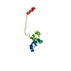
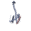
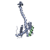
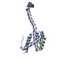
| Structure viewer | Molecule: MolmilJmol/JSmol |
|---|
- Downloads & links
-Download
| PDBx/mmCIF format | 1i2h.cif.gz | 43.2 KB | Display | PDBx/mmCIF format |
|---|---|---|---|---|
| PDB format | pdb1i2h.ent.gz | 28.9 KB | Display | PDB format |
| PDBx/mmJSON format | 1i2h.json.gz | Tree view | PDBx/mmJSON format | |
| Others |  Other downloads Other downloads |
-Validation report
| Arichive directory | https://data.pdbj.org/pub/pdb/validation_reports/i2/1i2hftp://data.pdbj.org/pub/pdb/validation_reports/i2/1i2h | HTTPS FTP |
|---|
-Related structure data
| Related structure data |  1ddwS S: Starting model for refinement |
|---|---|
| Similar structure data |


-Links
 PDBj
PDBj- Assembly
| Deposited unit | 
| ||||||||
|---|---|---|---|---|---|---|---|---|---|
| 1 |
| ||||||||
| Unit cell |
| ||||||||
| Components on special symmetry positions |
|
-Components
| #1: Protein | Mass: 18823.729 Da / Num. of mol.: 1 / Fragment: N-TERMINAL CONSERVED HOMER 1 REGION (CH1 DOMAIN) Source method: isolated from a genetically manipulated source Source: (gene. exp.)  |
|---|---|
| #2: Water | ChemComp-HOH /  Mass: 18.015 Da / Num. of mol.: 114 / Source method: isolated from a natural source / Formula: H2O Mass: 18.015 Da / Num. of mol.: 114 / Source method: isolated from a natural source / Formula: H2O |
-Experimental details
-Experiment
| Experiment | Method: X-RAY DIFFRACTION / Number of used crystals: 1 |
|---|
- Sample preparation
| Crystal | Density Matthews: 2.07 Å3/Da / Density % sol: 40.44 % | ||||||||||||||||||
|---|---|---|---|---|---|---|---|---|---|---|---|---|---|---|---|---|---|---|---|
| Crystal grow | Temperature: 293 K / Method: vapor diffusion, hanging drop / pH: 6.5 Details: sodium citrate, pH 6.5, VAPOR DIFFUSION, HANGING DROP, temperature 293K | ||||||||||||||||||
| Crystal grow | *PLUS | ||||||||||||||||||
| Components of the solutions | *PLUS
|
-Data collection
| Diffraction | Mean temperature: 100 K |
|---|---|
| Diffraction source | Source: SYNCHROTRON / Site: SPring-8  / Beamline: BL45XU / Wavelength: 1.02 Å / Beamline: BL45XU / Wavelength: 1.02 Å |
| Detector | Type: RIGAKU RAXIS V / Detector: IMAGE PLATE / Date: Nov 1, 2000 |
| Radiation | Monochromator: YALE MIRRORS / Protocol: SINGLE WAVELENGTH / Monochromatic (M) / Laue (L): M / Scattering type: x-ray |
| Radiation wavelength | Wavelength: 1.02 Å / Relative weight: 1 |
| Reflection | Resolution: 1.8→200 Å / Num. all: 15030 / Num. obs: 14936 / % possible obs: 99.4 % / Observed criterion σ(F): 1 / Observed criterion σ(I): 1 / Redundancy: 3.4 % / Biso Wilson estimate: 26.7 Å2 / Rmerge(I) obs: 0.066 / Net I/σ(I): 15 |
| Reflection shell | Resolution: 1.8→1.86 Å / Redundancy: 3.26 % / Rmerge(I) obs: 0.268 / % possible all: 96.7 |
| Reflection | *PLUS Highest resolution: 1.8 Å / Num. measured all: 50784 / Rmerge(I) obs: 0.066 |
| Reflection shell | *PLUS % possible obs: 96.7 % / Rmerge(I) obs: 0.268 |
- Processing
| Software |
| |||||||||||||||||||||||||
|---|---|---|---|---|---|---|---|---|---|---|---|---|---|---|---|---|---|---|---|---|---|---|---|---|---|---|
| Refinement | Method to determine structure: MOLECULAR REPLACEMENT Starting model: PDB ENTRY 1DDW Resolution: 1.8→19.09 Å / σ(F): 0 / σ(I): 0 / Stereochemistry target values: Engh & Huber
| |||||||||||||||||||||||||
| Refinement step | Cycle: LAST / Resolution: 1.8→19.09 Å
| |||||||||||||||||||||||||
| LS refinement shell | Resolution: 1.8→1.885 Å /
| |||||||||||||||||||||||||
| Refinement | *PLUS Lowest resolution: 19.09 Å / % reflection Rfree: 5 % / Rfactor all: 0.224 / Rfactor obs: 0.195 / Rfactor Rfree: 0.218 / Rfactor Rwork: 0.195 | |||||||||||||||||||||||||
| Solvent computation | *PLUS | |||||||||||||||||||||||||
| Displacement parameters | *PLUS | |||||||||||||||||||||||||
| Refine LS restraints | *PLUS
| |||||||||||||||||||||||||
| LS refinement shell | *PLUS Rfactor Rfree: 0.27 / Rfactor Rwork: 0.24 |
