+ データを開く
- 基本情報
基本情報
| 登録情報 | データベース: PDB / ID: 1hx6 | ||||||
|---|---|---|---|---|---|---|---|
| タイトル | P3, THE MAJOR COAT PROTEIN OF THE LIPID-CONTAINING BACTERIOPHAGE PRD1. | ||||||
 要素 要素 | MAJOR CAPSID PROTEIN | ||||||
 キーワード キーワード | VIRAL PROTEIN / bacteriophage PRD1 / coat protein / jelly roll / viral beta barrel | ||||||
| 機能・相同性 |  機能・相同性情報 機能・相同性情報 | ||||||
| 生物種 |   Enterobacteria phage PRD1 (ファージ) Enterobacteria phage PRD1 (ファージ) | ||||||
| 手法 |  X線回折 / X線回折 /  シンクロトロン / シンクロトロン /  分子置換 / 解像度: 1.65 Å 分子置換 / 解像度: 1.65 Å | ||||||
 データ登録者 データ登録者 | Benson, S.D. / Bamford, J.K.H. / Bamford, D.H. / Burnett, R.M. | ||||||
 引用 引用 |  ジャーナル: Acta Crystallogr D Biol Crystallogr / 年: 2002 ジャーナル: Acta Crystallogr D Biol Crystallogr / 年: 2002タイトル: The X-ray crystal structure of P3, the major coat protein of the lipid-containing bacteriophage PRD1, at 1.65 A resolution. 著者: Stacy D Benson / Jaana K H Bamford / Dennis H Bamford / Roger M Burnett /  要旨: P3 has been imaged with X-ray crystallography to reveal a trimeric molecule with strikingly similar characteristics to hexon, the major coat protein of adenovirus. The structure of native P3 has now ...P3 has been imaged with X-ray crystallography to reveal a trimeric molecule with strikingly similar characteristics to hexon, the major coat protein of adenovirus. The structure of native P3 has now been extended to 1.65 A resolution (R(work) = 19.0% and R(free) = 20.8%). The new high-resolution model shows that P3 forms crystals through hydrophobic patches solvated by 2-methyl-2,4-pentanediol molecules. It reveals details of how the molecule's high stability may be achieved through ordered solvent in addition to intra- and intersubunit interactions. Of particular importance is a 'puddle' at the top of the molecule containing a four-layer deep hydration shell that cross-links a complex structural feature formed by 'trimerization loops'. These loops also link subunits by extending over a neighbor to reach the third subunit in the trimer. As each subunit has two eight-stranded viral jelly rolls, the trimer has a pseudo-hexagonal shape to allow close packing in its 240 hexavalent capsid positions. Flexible regions in P3 facilitate these interactions within the capsid and with the underlying membrane. A selenometh-ionine P3 derivative, with which the structure was solved, has been refined to 2.2 A resolution (R(work) = 20.1% and R(free) = 22.8%). The derivatized molecule is essentially unchanged, although synchrotron radiation has the curious effect of causing it to rotate about its threefold axis. P3 is a second example of a trimeric 'double-barrel' protein that forms a stable building block with optimal shape for constructing a large icosahedral viral capsid. A major difference is that hexon has long variable loops that distinguish different adenovirus species. The short loops in P3 and the severe constraints of its various interactions explain why the PRD1 family has highly conserved coat proteins. #1:  ジャーナル: Cell(Cambridge,Mass.) / 年: 1999 ジャーナル: Cell(Cambridge,Mass.) / 年: 1999タイトル: Viral Evolution Revealed by Bacteriophage PRD1 and Human Adenovirus Coat Protein Structures 著者: Benson, S.D. / Bamford, J.K. / Bamford, D.H. / Burnett, R.M. #2:  ジャーナル: J.Mol.Biol. / 年: 1993 ジャーナル: J.Mol.Biol. / 年: 1993タイトル: Crystallization of the Major Coat Protein of PRD1, a Bacteriophage with an Internal Membrane 著者: Stewart, P.L. / Ghosh, S. / Bamford, D.H. / Burnett, R.M. #3:  ジャーナル: Virology / 年: 1990 ジャーナル: Virology / 年: 1990タイトル: Capsomer Proteins of Bacteriophage PRD1, a Bacterial Virus with a Membrane 著者: Bamford, J.K. / Bamford, D.H. #4:  ジャーナル: MOL.THER. / 年: 2000 ジャーナル: MOL.THER. / 年: 2000タイトル: Type-specific Epitope Locations Revealed by X-ray Crystallographic Study of Adenovirus Type 5 Hexon 著者: Rux, J.J. / Burnett, R.M. | ||||||
| 履歴 |
|
- 構造の表示
構造の表示
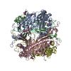
| 構造ビューア | 分子:  Molmil Molmil Jmol/JSmol Jmol/JSmol |
|---|
- ダウンロードとリンク
- ダウンロード
| PDBx/mmCIF形式 |  1hx6.cif.gz 1hx6.cif.gz | 254.6 KB | 表示 |  PDBx/mmCIF形式 PDBx/mmCIF形式 |
|---|---|---|---|---|
| PDB形式 |  pdb1hx6.ent.gz pdb1hx6.ent.gz | 206.5 KB | 表示 |  PDB形式 PDB形式 |
| PDBx/mmJSON形式 |  1hx6.json.gz 1hx6.json.gz | ツリー表示 |  PDBx/mmJSON形式 PDBx/mmJSON形式 | |
| その他 |  その他のダウンロード その他のダウンロード |
-検証レポート
| 文書・要旨 |  1hx6_validation.pdf.gz 1hx6_validation.pdf.gz | 478 KB | 表示 |  wwPDB検証レポート wwPDB検証レポート |
|---|---|---|---|---|
| 文書・詳細版 |  1hx6_full_validation.pdf.gz 1hx6_full_validation.pdf.gz | 498.9 KB | 表示 | |
| XML形式データ |  1hx6_validation.xml.gz 1hx6_validation.xml.gz | 50.2 KB | 表示 | |
| CIF形式データ |  1hx6_validation.cif.gz 1hx6_validation.cif.gz | 73.4 KB | 表示 | |
| アーカイブディレクトリ |  https://data.pdbj.org/pub/pdb/validation_reports/hx/1hx6 https://data.pdbj.org/pub/pdb/validation_reports/hx/1hx6 ftp://data.pdbj.org/pub/pdb/validation_reports/hx/1hx6 ftp://data.pdbj.org/pub/pdb/validation_reports/hx/1hx6 | HTTPS FTP |
-関連構造データ
- リンク
リンク
- 集合体
集合体
| 登録構造単位 | 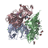
| ||||||||
|---|---|---|---|---|---|---|---|---|---|
| 1 |
| ||||||||
| 単位格子 |
|
- 要素
要素
| #1: タンパク質 | 分子量: 43346.219 Da / 分子数: 3 / 由来タイプ: 組換発現 由来: (組換発現)   Enterobacteria phage PRD1 (ファージ) Enterobacteria phage PRD1 (ファージ)属: Tectivirus / 発現宿主:  Salmonella typhimurium (サルモネラ菌) / 株 (発現宿主): DS88 / 参照: UniProt: P22535 Salmonella typhimurium (サルモネラ菌) / 株 (発現宿主): DS88 / 参照: UniProt: P22535#2: 化合物 | #3: 化合物 | ChemComp-NA / #4: 化合物 | ChemComp-MPD / ( #5: 水 | ChemComp-HOH / | |
|---|
-実験情報
-実験
| 実験 | 手法:  X線回折 / 使用した結晶の数: 1 X線回折 / 使用した結晶の数: 1 |
|---|
- 試料調製
試料調製
| 結晶 | マシュー密度: 3.48 Å3/Da / 溶媒含有率: 64.4 % |
|---|---|
| 結晶化 | 温度: 298 K / 手法: 蒸気拡散法, ハンギングドロップ法 / pH: 4.2 詳細: 30% MPD, 0.1 M sodium acetate, 0.2 M NaCl, pH 4.2, VAPOR DIFFUSION, HANGING DROP, temperature 298K |
-データ収集
| 回折 | 平均測定温度: 100 K |
|---|---|
| 放射光源 | 由来:  シンクロトロン / サイト: シンクロトロン / サイト:  NSLS NSLS  / ビームライン: X12C / 波長: 0.95 Å / ビームライン: X12C / 波長: 0.95 Å |
| 検出器 | タイプ: BRANDEIS / 検出器: CCD / 日付: 1997年6月11日 / 詳細: mirrors |
| 放射 | モノクロメーター: SI(III) / プロトコル: SINGLE WAVELENGTH / 単色(M)・ラウエ(L): M / 散乱光タイプ: x-ray |
| 放射波長 | 波長: 0.95 Å / 相対比: 1 |
| 反射 | 解像度: 1.6→87 Å / Num. obs: 195631 / % possible obs: 94.2 % / Observed criterion σ(I): 2 / 冗長度: 4.6 % / Biso Wilson estimate: 17.7 Å2 / Rsym value: 4.7 / Net I/σ(I): 20 |
| 反射 シェル | 解像度: 1.65→1.69 Å / 冗長度: 1.5 % / Num. unique all: 8530 / Rsym value: 40.3 / % possible all: 61.2 |
- 解析
解析
| ソフトウェア |
| ||||||||||||||||||||||||||||||||||||
|---|---|---|---|---|---|---|---|---|---|---|---|---|---|---|---|---|---|---|---|---|---|---|---|---|---|---|---|---|---|---|---|---|---|---|---|---|---|
| 精密化 | 構造決定の手法:  分子置換 分子置換開始モデル: selenomethionine P3 - PDB entry 1HQN 解像度: 1.65→53.45 Å / Rfactor Rfree error: 0.005 / Data cutoff high absF: 360397.3 / Data cutoff low absF: 0 / Isotropic thermal model: RESTRAINED / 交差検証法: THROUGHOUT / σ(F): 0 / 立体化学のターゲット値: Engh & Huber / 詳細: maximum likelihood
| ||||||||||||||||||||||||||||||||||||
| 溶媒の処理 | 溶媒モデル: FLAT MODEL / Bsol: 48.85 Å2 / ksol: 0.332 e/Å3 | ||||||||||||||||||||||||||||||||||||
| 原子変位パラメータ | Biso mean: 27 Å2
| ||||||||||||||||||||||||||||||||||||
| Refine analyze |
| ||||||||||||||||||||||||||||||||||||
| 精密化ステップ | サイクル: LAST / 解像度: 1.65→53.45 Å
| ||||||||||||||||||||||||||||||||||||
| 拘束条件 |
| ||||||||||||||||||||||||||||||||||||
| LS精密化 シェル | 解像度: 1.65→1.71 Å / Rfactor Rfree error: 0.026 / Total num. of bins used: 10
| ||||||||||||||||||||||||||||||||||||
| Xplor file |
|
 ムービー
ムービー コントローラー








 PDBj
PDBj


