 Movie
Movie Controller

[English] 日本語
 Yorodumi
- PDB-1hx6: P3, THE MAJOR COAT PROTEIN OF THE LIPID-CONTAINING BACTERIOPHAGE PRD1. -
+ Open data

- Basic information
| Entry | Database: PDB / ID: 1hx6 | ||||||
|---|---|---|---|---|---|---|---|
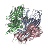
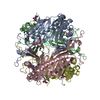
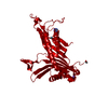
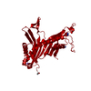
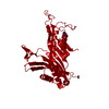
| Title | P3, THE MAJOR COAT PROTEIN OF THE LIPID-CONTAINING BACTERIOPHAGE PRD1. | ||||||
 Components Components | MAJOR CAPSID PROTEIN | ||||||
 Keywords Keywords | VIRAL PROTEIN / bacteriophage PRD1 / coat protein / jelly roll / viral beta barrel | ||||||
| Function / homology |  Function and homology information Function and homology information | ||||||
| Biological species |   Enterobacteria phage PRD1 (virus) Enterobacteria phage PRD1 (virus) | ||||||
| Method |  X-RAY DIFFRACTION / SYNCHROTRON / MOLECULAR REPLACEMENT / Resolution: 1.65 Å X-RAY DIFFRACTION / SYNCHROTRON / MOLECULAR REPLACEMENT / Resolution: 1.65 Å | ||||||
 Authors Authors | Benson, S.D. / Bamford, J.K.H. / Bamford, D.H. / Burnett, R.M. | ||||||
 Citation Citation | Journal: Acta Crystallogr D Biol Crystallogr / Year: 2002 Title: The X-ray crystal structure of P3, the major coat protein of the lipid-containing bacteriophage PRD1, at 1.65 A resolution. Authors: Stacy D Benson / Jaana K H Bamford / Dennis H Bamford / Roger M Burnett /  Abstract: P3 has been imaged with X-ray crystallography to reveal a trimeric molecule with strikingly similar characteristics to hexon, the major coat protein of adenovirus. The structure of native P3 has now ...P3 has been imaged with X-ray crystallography to reveal a trimeric molecule with strikingly similar characteristics to hexon, the major coat protein of adenovirus. The structure of native P3 has now been extended to 1.65 A resolution (R(work) = 19.0% and R(free) = 20.8%). The new high-resolution model shows that P3 forms crystals through hydrophobic patches solvated by 2-methyl-2,4-pentanediol molecules. It reveals details of how the molecule's high stability may be achieved through ordered solvent in addition to intra- and intersubunit interactions. Of particular importance is a 'puddle' at the top of the molecule containing a four-layer deep hydration shell that cross-links a complex structural feature formed by 'trimerization loops'. These loops also link subunits by extending over a neighbor to reach the third subunit in the trimer. As each subunit has two eight-stranded viral jelly rolls, the trimer has a pseudo-hexagonal shape to allow close packing in its 240 hexavalent capsid positions. Flexible regions in P3 facilitate these interactions within the capsid and with the underlying membrane. A selenometh-ionine P3 derivative, with which the structure was solved, has been refined to 2.2 A resolution (R(work) = 20.1% and R(free) = 22.8%). The derivatized molecule is essentially unchanged, although synchrotron radiation has the curious effect of causing it to rotate about its threefold axis. P3 is a second example of a trimeric 'double-barrel' protein that forms a stable building block with optimal shape for constructing a large icosahedral viral capsid. A major difference is that hexon has long variable loops that distinguish different adenovirus species. The short loops in P3 and the severe constraints of its various interactions explain why the PRD1 family has highly conserved coat proteins. #1: Journal: Cell(Cambridge,Mass.) / Year: 1999Title: Viral Evolution Revealed by Bacteriophage PRD1 and Human Adenovirus Coat Protein Structures Authors: Benson, S.D. / Bamford, J.K. / Bamford, D.H. / Burnett, R.M. #2: Journal: J.Mol.Biol. / Year: 1993Title: Crystallization of the Major Coat Protein of PRD1, a Bacteriophage with an Internal Membrane Authors: Stewart, P.L. / Ghosh, S. / Bamford, D.H. / Burnett, R.M. #3: Journal: Virology / Year: 1990Title: Capsomer Proteins of Bacteriophage PRD1, a Bacterial Virus with a Membrane Authors: Bamford, J.K. / Bamford, D.H. #4: Journal: MOL.THER. / Year: 2000Title: Type-specific Epitope Locations Revealed by X-ray Crystallographic Study of Adenovirus Type 5 Hexon Authors: Rux, J.J. / Burnett, R.M. | ||||||
| History |
|
- Structure visualization
| Structure viewer | Molecule: MolmilJmol/JSmol |
|---|
- Downloads & links
-Download
| PDBx/mmCIF format | 1hx6.cif.gz | 254.6 KB | Display | PDBx/mmCIF format |
|---|---|---|---|---|
| PDB format | pdb1hx6.ent.gz | 206.5 KB | Display | PDB format |
| PDBx/mmJSON format | 1hx6.json.gz | Tree view | PDBx/mmJSON format | |
| Others |  Other downloads Other downloads |
-Validation report
| Summary document | 1hx6_validation.pdf.gz | 478 KB | Display | wwPDB validaton report |
|---|---|---|---|---|
| Full document | 1hx6_full_validation.pdf.gz | 498.9 KB | Display | |
| Data in XML | 1hx6_validation.xml.gz | 50.2 KB | Display | |
| Data in CIF | 1hx6_validation.cif.gz | 73.4 KB | Display | |
| Arichive directory | https://data.pdbj.org/pub/pdb/validation_reports/hx/1hx6ftp://data.pdbj.org/pub/pdb/validation_reports/hx/1hx6 | HTTPS FTP |
-Related structure data
| Related structure data | 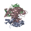 1hqnSC S: Starting model for refinement C: citing same article ( |
|---|---|
| Similar structure data |


-Links
 PDBj
PDBj

- Assembly
| Deposited unit | 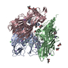
| ||||||||
|---|---|---|---|---|---|---|---|---|---|
| 1 |
| ||||||||
| Unit cell |
|
-Components
| #1: Protein | Mass: 43346.219 Da / Num. of mol.: 3 Source method: isolated from a genetically manipulated source Source: (gene. exp.) Enterobacteria phage PRD1 (virus) / Genus: Tectivirus / Production host:  Salmonella typhimurium (bacteria) / Strain (production host): DS88 / References: UniProt: P22535 Salmonella typhimurium (bacteria) / Strain (production host): DS88 / References: UniProt: P22535#2: Chemical |   Mass: 35.453 Da / Num. of mol.: 2 / Source method: obtained synthetically / Formula: Cl Mass: 35.453 Da / Num. of mol.: 2 / Source method: obtained synthetically / Formula: Cl#3: Chemical | ChemComp-NA /   Mass: 22.990 Da / Num. of mol.: 7 / Source method: obtained synthetically / Formula: Na Mass: 22.990 Da / Num. of mol.: 7 / Source method: obtained synthetically / Formula: Na#4: Chemical | ChemComp-MPD / (   Mass: 118.174 Da / Num. of mol.: 18 / Source method: obtained synthetically / Formula: C6H14O2 / Comment: precipitant*YM Mass: 118.174 Da / Num. of mol.: 18 / Source method: obtained synthetically / Formula: C6H14O2 / Comment: precipitant*YM#5: Water | ChemComp-HOH / |  Mass: 18.015 Da / Num. of mol.: 782 / Source method: isolated from a natural source / Formula: H2O Mass: 18.015 Da / Num. of mol.: 782 / Source method: isolated from a natural source / Formula: H2O |
|---|
-Experimental details
-Experiment
| Experiment | Method: X-RAY DIFFRACTION / Number of used crystals: 1 |
|---|
- Sample preparation
| Crystal | Density Matthews: 3.48 Å3/Da / Density % sol: 64.4 % |
|---|---|
| Crystal grow | Temperature: 298 K / Method: vapor diffusion, hanging drop / pH: 4.2 Details: 30% MPD, 0.1 M sodium acetate, 0.2 M NaCl, pH 4.2, VAPOR DIFFUSION, HANGING DROP, temperature 298K |
-Data collection
| Diffraction | Mean temperature: 100 K |
|---|---|
| Diffraction source | Source: SYNCHROTRON / Site: NSLS / Beamline: X12C / Wavelength: 0.95 Å |
| Detector | Type: BRANDEIS / Detector: CCD / Date: Jun 11, 1997 / Details: mirrors |
| Radiation | Monochromator: SI(III) / Protocol: SINGLE WAVELENGTH / Monochromatic (M) / Laue (L): M / Scattering type: x-ray |
| Radiation wavelength | Wavelength: 0.95 Å / Relative weight: 1 |
| Reflection | Resolution: 1.6→87 Å / Num. obs: 195631 / % possible obs: 94.2 % / Observed criterion σ(I): 2 / Redundancy: 4.6 % / Biso Wilson estimate: 17.7 Å2 / Rsym value: 4.7 / Net I/σ(I): 20 |
| Reflection shell | Resolution: 1.65→1.69 Å / Redundancy: 1.5 % / Num. unique all: 8530 / Rsym value: 40.3 / % possible all: 61.2 |
- Processing
| Software |
| ||||||||||||||||||||||||||||||||||||
|---|---|---|---|---|---|---|---|---|---|---|---|---|---|---|---|---|---|---|---|---|---|---|---|---|---|---|---|---|---|---|---|---|---|---|---|---|---|
| Refinement | Method to determine structure: MOLECULAR REPLACEMENT Starting model: selenomethionine P3 - PDB entry 1HQN Resolution: 1.65→53.45 Å / Rfactor Rfree error: 0.005 / Data cutoff high absF: 360397.3 / Data cutoff low absF: 0 / Isotropic thermal model: RESTRAINED / Cross valid method: THROUGHOUT / σ(F): 0 / Stereochemistry target values: Engh & Huber / Details: maximum likelihood
| ||||||||||||||||||||||||||||||||||||
| Solvent computation | Solvent model: FLAT MODEL / Bsol: 48.85 Å2 / ksol: 0.332 e/Å3 | ||||||||||||||||||||||||||||||||||||
| Displacement parameters | Biso mean: 27 Å2
| ||||||||||||||||||||||||||||||||||||
| Refine analyze |
| ||||||||||||||||||||||||||||||||||||
| Refinement step | Cycle: LAST / Resolution: 1.65→53.45 Å
| ||||||||||||||||||||||||||||||||||||
| Refine LS restraints |
| ||||||||||||||||||||||||||||||||||||
| LS refinement shell | Resolution: 1.65→1.71 Å / Rfactor Rfree error: 0.026 / Total num. of bins used: 10
| ||||||||||||||||||||||||||||||||||||
| Xplor file |
|
