 Movie
Movie Controller

[English] 日本語
 Yorodumi
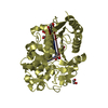
- PDB-1hrt: THE STRUCTURE OF A COMPLEX OF BOVINE ALPHA-THROMBIN AND RECOMBINA... -
+ Open data

- Basic information
| Entry | Database: PDB / ID: 1hrt | ||||||
|---|---|---|---|---|---|---|---|
| Title | THE STRUCTURE OF A COMPLEX OF BOVINE ALPHA-THROMBIN AND RECOMBINANT HIRUDIN AT 2.8 ANGSTROMS RESOLUTION | ||||||
 Components Components |
| ||||||
 Keywords Keywords | HYDROLASE/HYDROLASE INHIBITOR / SERINE PROTEINASE / HYDROLASE-HYDROLASE INHIBITOR COMPLEX | ||||||
| Function / homology |  Function and homology information Function and homology informationfibrinogen binding / thrombin / positive regulation of blood coagulation / protein polymerization / acute-phase response / serine-type endopeptidase inhibitor activity / platelet activation / toxin activity / serine-type endopeptidase activity / calcium ion binding ...fibrinogen binding / thrombin / positive regulation of blood coagulation / protein polymerization / acute-phase response / serine-type endopeptidase inhibitor activity / platelet activation / toxin activity / serine-type endopeptidase activity / calcium ion binding / proteolysis / : Similarity search - Function | ||||||
| Biological species |   Hirudo medicinalis (medicinal leech) Hirudo medicinalis (medicinal leech) | ||||||
| Method |  X-RAY DIFFRACTION / Resolution: 2.8 Å X-RAY DIFFRACTION / Resolution: 2.8 Å | ||||||
 Authors Authors | Vitali, J. / Edwards, B.F.P. | ||||||
 Citation Citation | Journal: J.Biol.Chem. / Year: 1992 Title: The structure of a complex of bovine alpha-thrombin and recombinant hirudin at 2.8-A resolution. Authors: Vitali, J. / Martin, P.D. / Malkowski, M.G. / Robertson, W.D. / Lazar, J.B. / Winant, R.C. / Johnson, P.H. / Edwards, B.F. | ||||||
| History |
|
- Structure visualization
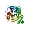
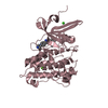
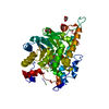
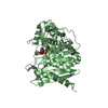
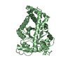
| Structure viewer | Molecule: MolmilJmol/JSmol |
|---|
- Downloads & links
-Download
| PDBx/mmCIF format | 1hrt.cif.gz | 86.6 KB | Display | PDBx/mmCIF format |
|---|---|---|---|---|
| PDB format | pdb1hrt.ent.gz | 64.8 KB | Display | PDB format |
| PDBx/mmJSON format | 1hrt.json.gz | Tree view | PDBx/mmJSON format | |
| Others |  Other downloads Other downloads |
-Validation report
| Arichive directory | https://data.pdbj.org/pub/pdb/validation_reports/hr/1hrtftp://data.pdbj.org/pub/pdb/validation_reports/hr/1hrt | HTTPS FTP |
|---|
-Related structure data
| Similar structure data |
|---|


-Links
 PDBj
PDBj


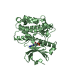
- Assembly
| Deposited unit | 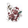
| ||||||||
|---|---|---|---|---|---|---|---|---|---|
| 1 |
| ||||||||
| Unit cell |
| ||||||||
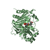
| Atom site foot note | 1: CIS PROLINE - PRO H 37 |
-Components
| #1: Protein/peptide | Mass: 5735.240 Da / Num. of mol.: 1 Source method: isolated from a genetically manipulated source Source: (gene. exp.) |
|---|---|
| #2: Protein | Mass: 29772.422 Da / Num. of mol.: 1 Source method: isolated from a genetically manipulated source Source: (gene. exp.) |
| #3: Protein | Mass: 6973.505 Da / Num. of mol.: 1 Source method: isolated from a genetically manipulated source Source: (gene. exp.) Hirudo medicinalis (medicinal leech) / Production host: unidentified (others) / References: UniProt: P01050 |
| #4: Water | ChemComp-HOH /  Mass: 18.015 Da / Num. of mol.: 129 / Source method: isolated from a natural source / Formula: H2O Mass: 18.015 Da / Num. of mol.: 129 / Source method: isolated from a natural source / Formula: H2O |
| Compound details | THROMBIN IS CLEAVED BETWEEN RESIDUES 15 AND 16. CHAIN INDICATOR *L* IS USED FOR RESIDUES 1H - 15 ...THROMBIN IS CLEAVED BETWEEN RESIDUES 15 AND 16. CHAIN INDICATOR *L* IS USED FOR RESIDUES 1H - 15 AND CHAIN INDICATOR *H* IS USED FOR RESIDUES 16 - 247. CHAIN INDICATOR *I* IS USED FOR HIRUDIN. THE COMPLEX CONSISTS OF ONE MOLECULE OF ALPHA-THROMBIN AND ONE MOLECULE OF HIRUDIN. |
| Has protein modification | Y |
-Experimental details
-Experiment
| Experiment | Method: X-RAY DIFFRACTION |
|---|
- Sample preparation
| Crystal | Density Matthews: 2.56 Å3/Da / Density % sol: 51.87 % | ||||||||||||||||||||||||||||||||||||
|---|---|---|---|---|---|---|---|---|---|---|---|---|---|---|---|---|---|---|---|---|---|---|---|---|---|---|---|---|---|---|---|---|---|---|---|---|---|
| Crystal grow | *PLUS Temperature: 22 ℃ / Method: vapor diffusion, hanging drop | ||||||||||||||||||||||||||||||||||||
| Components of the solutions | *PLUS
|
-Data collection
| Radiation | Scattering type: x-ray |
|---|---|
| Radiation wavelength | Relative weight: 1 |
| Reflection | *PLUS Highest resolution: 2.6 Å / Lowest resolution: 9999 Å / Num. obs: 7688 / Observed criterion σ(I): 1 / Num. measured all: 19008 / Rmerge(I) obs: 0.099 |
| Reflection shell | *PLUS Highest resolution: 2.6 Å / Lowest resolution: 2.8 Å / % possible obs: 13 % |
- Processing
| Software | Name: PROFFT / Classification: refinement | |||||||||||||||||||||||||||||||||||||||||||||||||||||||||||||||
|---|---|---|---|---|---|---|---|---|---|---|---|---|---|---|---|---|---|---|---|---|---|---|---|---|---|---|---|---|---|---|---|---|---|---|---|---|---|---|---|---|---|---|---|---|---|---|---|---|---|---|---|---|---|---|---|---|---|---|---|---|---|---|---|---|
| Refinement | Resolution: 2.8→7 Å / σ(F): 0 Details: SUBSEQUENT TO PUBLICATION, ADDITIONAL REFINEMENT WAS CARRIED OUT WITH PROFFT USING DAMPING FACTORS OF 0.7 FOR BOTH POSITIONAL AND THERMAL PARAMETERS. THE USE OF THIS DAMPING FACTOR COMPARED ...Details: SUBSEQUENT TO PUBLICATION, ADDITIONAL REFINEMENT WAS CARRIED OUT WITH PROFFT USING DAMPING FACTORS OF 0.7 FOR BOTH POSITIONAL AND THERMAL PARAMETERS. THE USE OF THIS DAMPING FACTOR COMPARED TO THE FULL SHIFTS APPLIED EARLIER REDUCED THE R VALUE FROM 0.161 REPORTED IN THE PAPER TO 0.155. THIS ENTRY CONTAINS THE REVISED COORDINATES. THESE COORDINATES GIVE THE SAME DISTANCES (WITHIN 0.2 ANGSTROMS) AS THOSE DISCUSSED IN THE PAPER.
| |||||||||||||||||||||||||||||||||||||||||||||||||||||||||||||||
| Refinement step | Cycle: LAST / Resolution: 2.8→7 Å
| |||||||||||||||||||||||||||||||||||||||||||||||||||||||||||||||
| Refine LS restraints |
| |||||||||||||||||||||||||||||||||||||||||||||||||||||||||||||||
| Software | *PLUS Name: PROFT / Classification: refinement | |||||||||||||||||||||||||||||||||||||||||||||||||||||||||||||||
| Refinement | *PLUS Highest resolution: 2.8 Å / Lowest resolution: 7 Å / Num. reflection all: 6958 / σ(F): 0 / Rfactor all: 0.155 | |||||||||||||||||||||||||||||||||||||||||||||||||||||||||||||||
| Solvent computation | *PLUS | |||||||||||||||||||||||||||||||||||||||||||||||||||||||||||||||
| Displacement parameters | *PLUS |
