Mass: 18.015 Da / Num. of mol.: 113 / Source method: isolated from a natural source / Formula: H2O
Source details
THE EXPRESSION PLASMID OF THE MUTANT HUMAN LYSOZYME, C77A, IN WHICH CYS 77 IS REPLACED BY ALA, WAS ...THE EXPRESSION PLASMID OF THE MUTANT HUMAN LYSOZYME, C77A, IN WHICH CYS 77 IS REPLACED BY ALA, WAS PREVIOUSLY DESCRIBED (TANIYAMA,Y., SEKO,C., AND KIKUCHI,M. (1990) J.BIOL.CHEM. 265, 16767-16771).
-
Experimental details
-
Experiment
Experiment
Method: X-RAY DIFFRACTION
-
Sample preparation
Crystal
Density Matthews: 1.97 Å3/Da / Density % sol: 37.59 %
Crystal grow
*PLUS
Temperature: 293 K / pH: 6 / Method: unknown / Details: using macroseeding
Components of the solutions
*PLUS
ID
Conc.
Common name
Crystal-ID
Sol-ID
Chemical formula
1
20mg/ml
protein
1
1
2
2.5M
1
2
NaCl
3
30mM
sodiumphosphate
1
2
-
Data collection
Radiation
Scattering type: x-ray
Radiation wavelength
Relative weight: 1
Reflection
Num. obs: 9501 / % possible obs: 84 %
Reflection
*PLUS
Highest resolution: 1.76 Å / Lowest resolution: 9999 Å / Num. measured all: 30257 / Rmerge(I) obs: 0.0667
-
Processing
Software
Name: PROLSQ / Classification: refinement
Refinement
Resolution: 1.8→5 Å /
Rfactor
Num. reflection
obs
0.125
8230
Displacement parameters
Biso mean: 18.4 Å2
Refinement step
Cycle: LAST / Resolution: 1.8→5 Å
Protein
Nucleic acid
Ligand
Solvent
Total
Num. atoms
1028
0
20
113
1161
Refine LS restraints
Refine-ID
Type
Dev ideal
Dev ideal target
X-RAY DIFFRACTION
p_bond_d
0.017
0.02
X-RAY DIFFRACTION
p_angle_d
0.04
0.04
X-RAY DIFFRACTION
p_angle_deg
X-RAY DIFFRACTION
p_planar_d
0.05
0.05
X-RAY DIFFRACTION
p_hb_or_metal_coord
X-RAY DIFFRACTION
p_mcbond_it
1.425
1.5
X-RAY DIFFRACTION
p_mcangle_it
2.313
2
X-RAY DIFFRACTION
p_scbond_it
2.283
2
X-RAY DIFFRACTION
p_scangle_it
3.552
2.5
X-RAY DIFFRACTION
p_plane_restr
0.014
0.02
X-RAY DIFFRACTION
p_chiral_restr
0.159
0.15
X-RAY DIFFRACTION
p_singtor_nbd
0.167
0.3
X-RAY DIFFRACTION
p_multtor_nbd
0.182
0.3
X-RAY DIFFRACTION
p_xhyhbond_nbd
0.221
0.3
X-RAY DIFFRACTION
p_xyhbond_nbd
X-RAY DIFFRACTION
p_planar_tor
2.6
3
X-RAY DIFFRACTION
p_staggered_tor
17.6
15
X-RAY DIFFRACTION
p_orthonormal_tor
20.2
20
X-RAY DIFFRACTION
p_transverse_tor
X-RAY DIFFRACTION
p_special_tor
+
About Yorodumi
-
News
-
Feb 9, 2022. New format data for meta-information of EMDB entries
New format data for meta-information of EMDB entries
Version 3 of the EMDB header file is now the official format.
The previous official version 1.9 will be removed from the archive.
In the structure databanks used in Yorodumi, some data are registered as the other names, "COVID-19 virus" and "2019-nCoV". Here are the details of the virus and the list of structure data.
Jan 31, 2019. EMDB accession codes are about to change! (news from PDBe EMDB page)
EMDB accession codes are about to change! (news from PDBe EMDB page)
The allocation of 4 digits for EMDB accession codes will soon come to an end. Whilst these codes will remain in use, new EMDB accession codes will include an additional digit and will expand incrementally as the available range of codes is exhausted. The current 4-digit format prefixed with “EMD-” (i.e. EMD-XXXX) will advance to a 5-digit format (i.e. EMD-XXXXX), and so on. It is currently estimated that the 4-digit codes will be depleted around Spring 2019, at which point the 5-digit format will come into force.
The EM Navigator/Yorodumi systems omit the EMD- prefix.
Related info.:Q: What is EMD? / ID/Accession-code notation in Yorodumi/EM Navigator
Yorodumi is a browser for structure data from EMDB, PDB, SASBDB, etc.
This page is also the successor to EM Navigator detail page, and also detail information page/front-end page for Omokage search.
The word "yorodu" (or yorozu) is an old Japanese word meaning "ten thousand". "mi" (miru) is to see.
Related info.:EMDB / PDB / SASBDB / Comparison of 3 databanks / Yorodumi Search / Aug 31, 2016. New EM Navigator & Yorodumi / Yorodumi Papers / Jmol/JSmol / Function and homology information / Changes in new EM Navigator and Yorodumi
 Movie
Movie Controller

 Yorodumi
 Open data
 Basic information
 Components
 Keywords
 Function and homology information
 Homo sapiens (human)
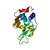
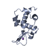
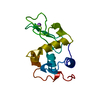
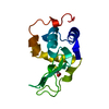
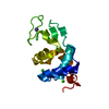
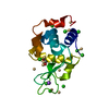
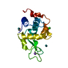
 X-RAY DIFFRACTION / Resolution: 1.8 Å
  Authors
 Citation
 Structure visualization
 Downloads & links
 Other downloads


 PDBj
PDBj


 Assembly

 Mass: 307.323 Da / Num. of mol.: 1 / Source method: obtained synthetically / Formula: C10H17N3O6S
 Mass: 18.015 Da / Num. of mol.: 113 / Source method: isolated from a natural source / Formula: H2O
 Sample preparation
 Processing