 Movie
Movie Controller

[English] 日本語
 Yorodumi
- PDB-1hhp: THE THREE-DIMENSIONAL STRUCTURE OF THE ASPARTYL PROTEASE FROM THE... -
+ Open data

- Basic information
| Entry | Database: PDB / ID: 1hhp | ||||||
|---|---|---|---|---|---|---|---|
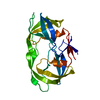
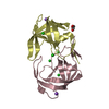
| Title | THE THREE-DIMENSIONAL STRUCTURE OF THE ASPARTYL PROTEASE FROM THE HIV-1 ISOLATE BRU | ||||||
 Components Components | UNLIGANDED HIV-1 PROTEASE | ||||||
 Keywords Keywords | HYDROLASE(ACID PROTEINASE) | ||||||
| Function / homology |  Function and homology information Function and homology informationHIV-1 retropepsin / symbiont-mediated activation of host apoptosis / retroviral ribonuclease H / exoribonuclease H / exoribonuclease H activity / DNA integration / viral genome integration into host DNA / establishment of integrated proviral latency / RNA-directed DNA polymerase / RNA stem-loop binding ...HIV-1 retropepsin / symbiont-mediated activation of host apoptosis / retroviral ribonuclease H / exoribonuclease H / exoribonuclease H activity / DNA integration / viral genome integration into host DNA / establishment of integrated proviral latency / RNA-directed DNA polymerase / RNA stem-loop binding / viral penetration into host nucleus / host multivesicular body / RNA-directed DNA polymerase activity / RNA-DNA hybrid ribonuclease activity / Transferases; Transferring phosphorus-containing groups; Nucleotidyltransferases / host cell / viral nucleocapsid / DNA recombination / DNA-directed DNA polymerase / aspartic-type endopeptidase activity / Hydrolases; Acting on ester bonds / DNA-directed DNA polymerase activity / symbiont-mediated suppression of host gene expression / viral translational frameshifting / symbiont entry into host cell / lipid binding / host cell nucleus / host cell plasma membrane / virion membrane / structural molecule activity / proteolysis / DNA binding / zinc ion binding Similarity search - Function | ||||||
| Biological species |   Human immunodeficiency virus type 1 Human immunodeficiency virus type 1 | ||||||
| Method |  X-RAY DIFFRACTION / Resolution: 2.7 Å X-RAY DIFFRACTION / Resolution: 2.7 Å | ||||||
 Authors Authors | Spinelli, S. / Alzari, P.M. | ||||||
 Citation Citation | Journal: Biochimie / Year: 1991 Title: The three-dimensional structure of the aspartyl protease from the HIV-1 isolate BRU. Authors: Spinelli, S. / Liu, Q.Z. / Alzari, P.M. / Hirel, P.H. / Poljak, R.J. | ||||||
| History |
|
- Structure visualization
| Structure viewer | Molecule: MolmilJmol/JSmol |
|---|
- Downloads & links
-Download
| PDBx/mmCIF format | 1hhp.cif.gz | 29.8 KB | Display | PDBx/mmCIF format |
|---|---|---|---|---|
| PDB format | pdb1hhp.ent.gz | 19.8 KB | Display | PDB format |
| PDBx/mmJSON format | 1hhp.json.gz | Tree view | PDBx/mmJSON format | |
| Others |  Other downloads Other downloads |
-Validation report
| Arichive directory | https://data.pdbj.org/pub/pdb/validation_reports/hh/1hhpftp://data.pdbj.org/pub/pdb/validation_reports/hh/1hhp | HTTPS FTP |
|---|
-Related structure data
| Similar structure data |
|---|


-Links
 PDBj
PDBj


- Assembly
| Deposited unit | 
| ||||||||
|---|---|---|---|---|---|---|---|---|---|
| 1 | 
| ||||||||
| Unit cell |
|
-Components
| #1: Protein | Mass: 10803.756 Da / Num. of mol.: 1 Source method: isolated from a genetically manipulated source Source: (gene. exp.) Human immunodeficiency virus type 1 (BRU ISOLATE)Genus: Lentivirus / Species: Human immunodeficiency virus 1 / Production host:  |
|---|
-Experimental details
-Experiment
| Experiment | Method: X-RAY DIFFRACTION |
|---|
- Sample preparation
| Crystal | Density Matthews: 3.11 Å3/Da / Density % sol: 60.48 % | ||||||||||||||||||||||||||||||
|---|---|---|---|---|---|---|---|---|---|---|---|---|---|---|---|---|---|---|---|---|---|---|---|---|---|---|---|---|---|---|---|
| Crystal grow | *PLUS Temperature: 4 ℃ / pH: 7 / Method: vapor diffusion, hanging drop | ||||||||||||||||||||||||||||||
| Components of the solutions | *PLUS
|
-Data collection
| Radiation | Scattering type: x-ray |
|---|---|
| Radiation wavelength | Relative weight: 1 |
| Reflection | *PLUS Highest resolution: 2.7 Å / Lowest resolution: 6 Å / Num. obs: 3549 / % possible obs: 93 % / Observed criterion σ(F): 2 / Num. measured all: 3901 / Rmerge(I) obs: 0.098 |
- Processing
| Software |
| ||||||||||||
|---|---|---|---|---|---|---|---|---|---|---|---|---|---|
| Refinement | Rfactor Rwork: 0.19 / Highest resolution: 2.7 Å | ||||||||||||
| Refinement step | Cycle: LAST / Highest resolution: 2.7 Å
| ||||||||||||
| Refine LS restraints |
| ||||||||||||
| Software | *PLUS Name: X-PLOR / Classification: refinement | ||||||||||||
| Refinement | *PLUS Lowest resolution: 6 Å / Num. reflection obs: 3549 / σ(F): 2 / Rfactor obs: 0.19 | ||||||||||||
| Solvent computation | *PLUS | ||||||||||||
| Displacement parameters | *PLUS Biso mean: 31 Å2 | ||||||||||||
| Refine LS restraints | *PLUS
|
