 Movie
Movie Controller

[English] 日本語
 Yorodumi
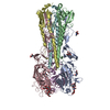
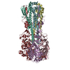
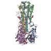
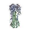
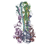
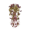
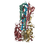
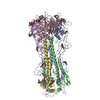
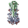
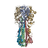
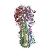
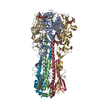
- PDB-1hgh: BINDING OF INFLUENZA VIRUS HEMAGGLUTININ TO ANALOGS OF ITS CELL-S... -
+ Open data

- Basic information
| Entry | Database: PDB / ID: 1hgh | |||||||||
|---|---|---|---|---|---|---|---|---|---|---|
| Title | BINDING OF INFLUENZA VIRUS HEMAGGLUTININ TO ANALOGS OF ITS CELL-SURFACE RECEPTOR, SIALIC ACID: ANALYSIS BY PROTON NUCLEAR MAGNETIC RESONANCE SPECTROSCOPY AND X-RAY CRYSTALLOGRAPHY | |||||||||
 Components Components | (HEMAGGLUTININ, CHAIN ...) x 2 | |||||||||
 Keywords Keywords | VIRAL PROTEIN / INFLUENZA VIRUS HEMAGGLUTININ | |||||||||
| Function / homology |  Function and homology information Function and homology informationviral budding from plasma membrane / clathrin-dependent endocytosis of virus by host cell / host cell surface receptor binding / fusion of virus membrane with host plasma membrane / fusion of virus membrane with host endosome membrane / viral envelope / virion attachment to host cell / host cell plasma membrane / virion membrane / membrane Similarity search - Function | |||||||||
| Biological species |   Influenza A virus Influenza A virus | |||||||||
| Method |  X-RAY DIFFRACTION / Resolution: 2.7 Å X-RAY DIFFRACTION / Resolution: 2.7 Å | |||||||||
 Authors Authors | Sauter, N.K. / Hanson, J.E. / Glick, G.D. / Brown, J.H. / Crowther, R.L. / Park, S.-J. / Skehel, J.J. / Wiley, D.C. | |||||||||
 Citation Citation | Journal: Biochemistry / Year: 1992 Title: Binding of influenza virus hemagglutinin to analogs of its cell-surface receptor, sialic acid: analysis by proton nuclear magnetic resonance spectroscopy and X-ray crystallography. Authors: Sauter, N.K. / Hanson, J.E. / Glick, G.D. / Brown, J.H. / Crowther, R.L. / Park, S.J. / Skehel, J.J. / Wiley, D.C. #1: Journal: Proc.Natl.Acad.Sci.USA / Year: 1992Title: Crystallographic Detection of a Second Ligand Binding Site in Influenza Virus Hemagglutinin Authors: Sauter, N.K. / Glick, G.D. / Crowther, R.L. / Park, S.-J. / Eisen, M.B. / Skehel, J.J. / Knowles, J.R. / Wiley, D.C. #2: Journal: J.Mol.Biol. / Year: 1990Title: Refinement of the Influenza Virus Hemagglutinin by Simulated Annealing Authors: Weis, W.I. / Brunger, A.T. / Skehel, J.J. / Wiley, D.C. #3: Journal: Nature / Year: 1988Title: Structure of the Influenza Virus Hemagglutinin Complexed with its Receptor, Sialic Acid Authors: Weis, W.I. / Brown, J.H. / Cusack, S. / Paulson, J.C. / Skehel, J.J. / Wiley, D.C. #4: Journal: Acta Crystallogr.,Sect.B / Year: 1986Title: The Refinement of the Hemagglutinin Membrane Glycoprotein of Influenza Virus Authors: Knossow, M. / Lewis, M. / Rees, D. / Wilson, I.A. / Skehel, J.J. / Wiley, D.C. #5: Journal: Nature / Year: 1984Title: Three-Dimensional Structure of an Antigenic Mutant of the Influenza Virus Hemagglutinin Authors: Knossow, M. / Daniels, R.S. / Douglas, A.R. / Skehel, J.J. / Wiley, D.C. #6: Journal: Nature / Year: 1981Title: Structure of the Hemagglutinin Membrane Glycoprotein of Influenza Virus at 3 Angstroms Resolution Authors: Wilson, I.A. / Skehel, J.J. / Wiley, D.C. #7: Journal: Nature / Year: 1981Title: Structural Identification of the Antibody-Binding Sites of Hong Kong Influenza Hemagglutinin and Their Involvement in Antigenic Variation Authors: Wiley, D.C. / Wilson, I.A. / Skehel, J.J. #8: Journal: J.Mol.Biol. / Year: 1977Title: Crystallization and X-Ray Diffraction Studies on the Hemagglutinin Glycoprotein from the Membrane of Influenza Virus Authors: Wiley, D.C. / Skehel, J.J. | |||||||||
| History |
|
- Structure visualization
| Structure viewer | Molecule: MolmilJmol/JSmol |
|---|
- Downloads & links
-Download
| PDBx/mmCIF format | 1hgh.cif.gz | 376.1 KB | Display | PDBx/mmCIF format |
|---|---|---|---|---|
| PDB format | pdb1hgh.ent.gz | 312.4 KB | Display | PDB format |
| PDBx/mmJSON format | 1hgh.json.gz | Tree view | PDBx/mmJSON format | |
| Others |  Other downloads Other downloads |
-Validation report
| Arichive directory | https://data.pdbj.org/pub/pdb/validation_reports/hg/1hghftp://data.pdbj.org/pub/pdb/validation_reports/hg/1hgh | HTTPS FTP |
|---|
-Related structure data
| Related structure data | 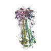 1hgdC  1hgeC  1hgfC 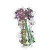 1hggC  1hgiC  1hgjC C: citing same article ( |
|---|---|
| Similar structure data |

-Links
 PDBj
PDBj


- Assembly
| Deposited unit | 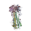
| ||||||||||||
|---|---|---|---|---|---|---|---|---|---|---|---|---|---|
| 1 |
| ||||||||||||
| Unit cell |
| ||||||||||||
| Atom site foot note | 1: DISORDERED RESIDUES WITH LITTLE OR NO VISIBLE ELECTRON DENSITY: CHAIN A: 1 - 8, 326 - 328 B: 58, 172 - 175 CHAIN C: 1 - 8, 328 D: 58, 172 - 175 CHAIN E: 1 - 8, 327 - 328 F: 58, 172 - 175 2: CIS PROLINE - PRO A 55 / 3: CIS PROLINE - PRO C 55 / 4: CIS PROLINE - PRO E 55 | ||||||||||||
| Noncrystallographic symmetry (NCS) | NCS oper:
| ||||||||||||
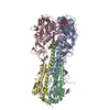
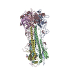
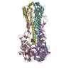
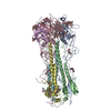
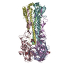
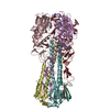
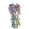
| Details | THERE IS ONE TRIMERIC HEMAGGLUTININ MOLECULE IN THE ASYMMETRIC UNIT, WITH THE MONOMERS RELATED TO EACH OTHER BY A NON-CRYSTALLOGRAPHIC THREE-FOLD AXIS. EACH MONOMER CONSISTS OF TWO CHAINS DESIGNATED HA1 AND HA2. HA1 AND HA2 OF MONOMER 1 ARE ASSIGNED CHAIN INDICATORS A AND B, RESPECTIVELY; HA1 AND HA2 OF MONOMER 2 ARE ASSIGNED CHAIN INDICATORS C AND D; AND HA1 AND HA2 OF MONOMER 3 ARE ASSIGNED CHAIN INDICATORS E AND F. LIGAND, N-LINKED CARBOHYDRATE, AND WATER MOLECULES ARE ASSIGNED SEPARATE CHAIN INDICATORS, ONE FOR EACH MONOMER: CHAIN G CONTAINS LIGAND, CARBOHYDRATE, AND WATER MOLECULES FOR MONOMER 1; CHAIN H CONTAINS LIGAND, CARBOHYDRATE, AND WATER MOLECULES FOR MONOMER 2; AND CHAIN I CONTAINS LIGAND, CARBOHYDRATE, AND WATER MOLECULES FOR MONOMER 3. CHAIN J CONTAINS THREE WATER MOLECULES BOUND IN CRYSTAL CONTACTS. IN THE VIRUS, CHAIN HA1 CONSISTS OF 328 RESIDUES AND CHAIN HA2 CONSISTS OF 220 RESIDUES. HEMAGGLUTININ MAY BE SOLUBILIZED FROM THE VIRAL MEMBRANE BY BROMELAIN DIGESTION, WHICH REMOVES THE C-TERMINAL HYDROPHOBIC (ANCHORING) DOMAIN FROM CHAIN HA2. AFTER BROMELAIN DIGESTION CHAIN HA2 CONSISTS OF 175 RESIDUES, AS PRESENTED IN THIS ENTRY. THE LIGAND, N-LINKED CARBOHYDRATE, AND WATER MOLECULES ASSOCIATED WITH EACH MONOMER ARE PRESENTED IMMEDIATELY FOLLOWING CHAIN HA2 OF THAT MONOMER AND HAVE BEEN ASSIGNED THE CHAIN INDICATORS G, H, AND I. THE TRANSFORMATION PRESENTED AS *MTRIX 1* BELOW WILL YIELD APPROXIMATE COORDINATES FOR MONOMER 2 (CHAINS C AND D) WHEN APPLIED TO MONOMER 1 (CHAINS A AND B). THE TRANSFORMATION PRESENTED AS *MTRIX 2* BELOW WILL YIELD APPROXIMATE COORDINATES FOR MONOMER 3 (CHAINS E AND F) WHEN APPLIED TO MONOMER 1 (CHAINS A AND B). THE TRANSFORMATIONS ARE DERIVED FROM THE POSITION OF THE NON-CRYSTALLOGRAPHIC THREE-FOLD SYMMETRY AXIS USED IN THE EARLY STAGES OF REFINEMENT WHEN STRICT THREE-FOLD SYMMETRY WAS IMPOSED ON THE STRUCTURE. THIS AXIS IS ALSO USED IN AVERAGING ELECTRON DENSITY MAPS. |
-Components
-HEMAGGLUTININ, CHAIN ... , 2 types, 6 molecules ACEBDF
| #1: Protein | Mass: 36065.457 Da / Num. of mol.: 3 Source method: isolated from a genetically manipulated source Source: (gene. exp.) Influenza A virus / Genus: Influenzavirus A / Strain: (A/X-31(H3N2)) / References: UniProt: P03437, UniProt: P03438*PLUS#2: Protein | Mass: 20212.350 Da / Num. of mol.: 3 Source method: isolated from a genetically manipulated source Source: (gene. exp.) Influenza A virus / Genus: Influenzavirus A / Strain: (A/X-31(H3N2)) / References: UniProt: P03437, UniProt: P03438*PLUS |
|---|
-Sugars , 3 types, 21 molecules 
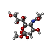
| #3: Polysaccharide | Source method: isolated from a genetically manipulated source #4: Sugar | ChemComp-NAG /  Type: D-saccharide, beta linking / Mass: 221.208 Da / Num. of mol.: 12 Type: D-saccharide, beta linking / Mass: 221.208 Da / Num. of mol.: 12Source method: isolated from a genetically manipulated source Formula: C8H15NO6 #5: Sugar | ChemComp-MNA /  Type: D-saccharide / Mass: 323.296 Da / Num. of mol.: 6 / Source method: obtained synthetically / Formula: C12H21NO9 Type: D-saccharide / Mass: 323.296 Da / Num. of mol.: 6 / Source method: obtained synthetically / Formula: C12H21NO9 |
|---|
-Non-polymers , 1 types, 72 molecules 
| #6: Water | ChemComp-HOH / Mass: 18.015 Da / Num. of mol.: 72 / Source method: isolated from a natural source / Formula: H2O |
|---|
-Details
| Has protein modification | Y |
|---|---|
| Nonpolymer details | SINCE THE ELECTRON DENSITY DOES NOT CLEARLY ESTABLISH THE CONFORMATION OF THE GLYCOSIDIC SIDE CHAIN ...SINCE THE ELECTRON DENSITY DOES NOT CLEARLY ESTABLISH THE CONFORMATI |
-Experimental details
-Experiment
| Experiment | Method: X-RAY DIFFRACTION |
|---|
- Sample preparation
| Crystal grow | *PLUS Method: other / Details: NMR |
|---|
-Data collection
| Radiation | Scattering type: x-ray |
|---|---|
| Radiation wavelength | Relative weight: 1 |
| Reflection | *PLUS Num. obs: 85146 / Num. measured all: 157204 / Rmerge(I) obs: 0.087 |
- Processing
| Software |
| ||||||||||||||||||||||||||||||||||||||||||||||||||||||||||||
|---|---|---|---|---|---|---|---|---|---|---|---|---|---|---|---|---|---|---|---|---|---|---|---|---|---|---|---|---|---|---|---|---|---|---|---|---|---|---|---|---|---|---|---|---|---|---|---|---|---|---|---|---|---|---|---|---|---|---|---|---|---|
| Refinement | Resolution: 2.7→7 Å / Rfactor Rwork: 0.226 / Rfactor obs: 0.226 | ||||||||||||||||||||||||||||||||||||||||||||||||||||||||||||
| Refinement step | Cycle: LAST / Resolution: 2.7→7 Å
| ||||||||||||||||||||||||||||||||||||||||||||||||||||||||||||
| Refine LS restraints |
| ||||||||||||||||||||||||||||||||||||||||||||||||||||||||||||
| Refinement | *PLUS Highest resolution: 2.7 Å / Lowest resolution: 7 Å / Num. reflection obs: 73501 / Rfactor obs: 0.226 | ||||||||||||||||||||||||||||||||||||||||||||||||||||||||||||
| Solvent computation | *PLUS | ||||||||||||||||||||||||||||||||||||||||||||||||||||||||||||
| Displacement parameters | *PLUS | ||||||||||||||||||||||||||||||||||||||||||||||||||||||||||||
| Refine LS restraints | *PLUS Type: x_angle_d / Dev ideal: 2.9 |
