 Movie
Movie Controller

[English] 日本語
 Yorodumi
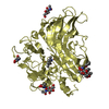
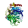
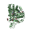
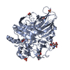
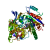
- PDB-1gyc: CRYSTAL STRUCTURE DETERMINATION AT ROOM TEMPERATURE OF A LACCASE ... -
+ Open data

- Basic information
| Entry | Database: PDB / ID: 1gyc | |||||||||
|---|---|---|---|---|---|---|---|---|---|---|
| Title | CRYSTAL STRUCTURE DETERMINATION AT ROOM TEMPERATURE OF A LACCASE FROM TRAMETES VERSICOLOR IN ITS OXIDISED FORM CONTAINING A FULL COMPLEMENT OF COPPER IONS | |||||||||
 Components Components | LACCASE 2 | |||||||||
 Keywords Keywords | OXIDOREDUCTASE / LACCASE / DIPHENOL OXIDASE / LIGNIN DEGRADATION | |||||||||
| Function / homology |  Function and homology information Function and homology informationlignin catabolic process / hydroquinone:oxygen oxidoreductase activity / laccase / copper ion binding / extracellular region Similarity search - Function | |||||||||
| Biological species |  TRAMETES VERSICOLOR (turkey-tail fungus) TRAMETES VERSICOLOR (turkey-tail fungus) | |||||||||
| Method |  X-RAY DIFFRACTION / SYNCHROTRON / MOLECULAR REPLACEMENT / Resolution: 1.9 Å X-RAY DIFFRACTION / SYNCHROTRON / MOLECULAR REPLACEMENT / Resolution: 1.9 Å | |||||||||
 Authors Authors | Choinowski, T. / Antorini, M. / Piontek, K. | |||||||||
 Citation Citation | Journal: J.Biol.Chem. / Year: 2002 Title: Crystal Structure of a Laccase from the Fungus Trametes Versicolor at 1.90-A Resolution Containing a Full Complement of Coppers. Authors: Piontek, K. / Antorini, M. / Choinowski, T. #1: Journal: Biochim.Biophys.Acta / Year: 2002 Title: Purificcation, Crystallisation and X-Ray Diffraction Study of Fully Functional Laccases from Two Ligninolytic Fungi Authors: Antorini, M. / Herpoel-Gimbertchoinowski, I. / Sigoillot-C, J. / Asther, M. / Winterhalter, K. | |||||||||
| History |
| |||||||||
| Remark 700 | SHEET THE SHEET STRUCTURE OF THIS MOLECULE IS BIFURCATED. IN ORDER TO REPRESENT THIS FEATURE IN ... SHEET THE SHEET STRUCTURE OF THIS MOLECULE IS BIFURCATED. IN ORDER TO REPRESENT THIS FEATURE IN THE SHEET RECORDS BELOW, TWO SHEETS ARE DEFINED. |
- Structure visualization
| Structure viewer | Molecule: MolmilJmol/JSmol |
|---|
- Downloads & links
-Download
| PDBx/mmCIF format | 1gyc.cif.gz | 129.1 KB | Display | PDBx/mmCIF format |
|---|---|---|---|---|
| PDB format | pdb1gyc.ent.gz | 98.6 KB | Display | PDB format |
| PDBx/mmJSON format | 1gyc.json.gz | Tree view | PDBx/mmJSON format | |
| Others |  Other downloads Other downloads |
-Validation report
| Arichive directory | https://data.pdbj.org/pub/pdb/validation_reports/gy/1gycftp://data.pdbj.org/pub/pdb/validation_reports/gy/1gyc | HTTPS FTP |
|---|
-Related structure data
| Related structure data |  1a65S S: Starting model for refinement |
|---|---|
| Similar structure data |


-Links
 PDBj
PDBj
- Assembly
| Deposited unit | 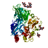
| ||||||||
|---|---|---|---|---|---|---|---|---|---|
| 1 |
| ||||||||
| Unit cell |
|
-Components
-Protein , 1 types, 1 molecules A
| #1: Protein | Mass: 53677.891 Da / Num. of mol.: 1 / Source method: isolated from a natural source / Source: (natural) TRAMETES VERSICOLOR (turkey-tail fungus) / References: UniProt: Q12718, laccase |
|---|
-Sugars , 2 types, 6 molecules 
| #2: Polysaccharide | Source method: isolated from a genetically manipulated source #4: Sugar |  Type: D-saccharide, beta linking / Mass: 221.208 Da / Num. of mol.: 3 Type: D-saccharide, beta linking / Mass: 221.208 Da / Num. of mol.: 3Source method: isolated from a genetically manipulated source Formula: C8H15NO6 |
|---|
-Non-polymers , 3 types, 596 molecules 


| #3: Chemical | ChemComp-CU /  Mass: 63.546 Da / Num. of mol.: 4 / Source method: obtained synthetically / Formula: Cu Mass: 63.546 Da / Num. of mol.: 4 / Source method: obtained synthetically / Formula: Cu#5: Chemical |  Mass: 60.095 Da / Num. of mol.: 2 / Source method: obtained synthetically / Formula: C3H8O Mass: 60.095 Da / Num. of mol.: 2 / Source method: obtained synthetically / Formula: C3H8O#6: Water | ChemComp-HOH / | Mass: 18.015 Da / Num. of mol.: 590 / Source method: isolated from a natural source / Formula: H2O |
|---|
-Details
| Has protein modification | Y |
|---|---|
| Sequence details | SWISS-PROT ENTRY Q12178 HAS THE CLOSEST AGREEMENT WITH THE SEQUENCE OF THE CRYSTALLIZED STRUCTURE. ...SWISS-PROT ENTRY Q12178 HAS THE CLOSEST AGREEMENT WITH THE SEQUENCE OF THE CRYSTALLIZ |
-Experimental details
-Experiment
| Experiment | Method: X-RAY DIFFRACTION / Number of used crystals: 1 |
|---|
- Sample preparation
| Crystal | Density Matthews: 2.61 Å3/Da / Density % sol: 52.53 % | ||||||||||||||||||||||||||||||||||||
|---|---|---|---|---|---|---|---|---|---|---|---|---|---|---|---|---|---|---|---|---|---|---|---|---|---|---|---|---|---|---|---|---|---|---|---|---|---|
| Crystal grow | pH: 5.6 Details: 20% (W/V) PEG8000, 20% (V/V) ISOPROPANOL, 100 MM SODIUM CITRATE PH 5.6 | ||||||||||||||||||||||||||||||||||||
| Crystal grow | *PLUS Method: vapor diffusion, hanging dropDetails: Antorini, M., (2002) Biochim.Biophys.Acta, 1594, 109. | ||||||||||||||||||||||||||||||||||||
| Components of the solutions | *PLUS
|
-Data collection
| Diffraction | Mean temperature: 287 K |
|---|---|
| Diffraction source | Source: SYNCHROTRON / Site: EMBL/DESY, HAMBURG  / Beamline: BW7B / Wavelength: 0.847 / Beamline: BW7B / Wavelength: 0.847 |
| Detector | Type: MAR scanner 345 mm plate / Detector: IMAGE PLATE / Date: Apr 15, 1999 |
| Radiation | Protocol: SINGLE WAVELENGTH / Monochromatic (M) / Laue (L): M / Scattering type: x-ray |
| Radiation wavelength | Wavelength: 0.847 Å / Relative weight: 1 |
| Reflection | Resolution: 1.9→20 Å / Num. obs: 52154 / % possible obs: 99.4 % / Redundancy: 4.4 % / Rmerge(I) obs: 0.063 / Net I/σ(I): 15.1 |
| Reflection | *PLUS Highest resolution: 1.9 Å / Lowest resolution: 20 Å / % possible obs: 99.7 % |
| Reflection shell | *PLUS Highest resolution: 1.9 Å / Lowest resolution: 1.97 Å / Rmerge(I) obs: 0.38 |
- Processing
| Software |
| ||||||||||||||||||||
|---|---|---|---|---|---|---|---|---|---|---|---|---|---|---|---|---|---|---|---|---|---|
| Refinement | Method to determine structure: MOLECULAR REPLACEMENT Starting model: 1A65 Resolution: 1.9→17.9 Å / Details: NONE
| ||||||||||||||||||||
| Refinement step | Cycle: LAST / Resolution: 1.9→17.9 Å
| ||||||||||||||||||||
| Refinement | *PLUS Lowest resolution: 20 Å / % reflection Rfree: 5 % / Rfactor Rfree: 0.209 / Rfactor Rwork: 0.168 | ||||||||||||||||||||
| Solvent computation | *PLUS | ||||||||||||||||||||
| Displacement parameters | *PLUS | ||||||||||||||||||||
| Refine LS restraints | *PLUS
| ||||||||||||||||||||
| LS refinement shell | *PLUS Highest resolution: 1.9 Å / Lowest resolution: 2.02 Å / Rfactor Rfree: 0.257 / Rfactor Rwork: 0.205 / Total num. of bins used: 15 |
