 Movie
Movie Controller


+ Open data

- Basic information
| Entry | Database: PDB / ID: 1gqa | ||||||
|---|---|---|---|---|---|---|---|
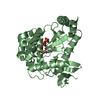
| Title | Cytochrome c' from Rhodobacter Spheriodes | ||||||
 Components Components | CYTOCHROME C' | ||||||
 Keywords Keywords | ELECTRON TRANSPORT / HEME | ||||||
| Function / homology |  Function and homology information Function and homology informationelectron transport chain / periplasmic space / electron transfer activity / iron ion binding / heme binding Similarity search - Function | ||||||
| Biological species |  RHODOBACTER SPHAEROIDES (bacteria) RHODOBACTER SPHAEROIDES (bacteria) | ||||||
| Method |  X-RAY DIFFRACTION / MOLECULAR REPLACEMENT / Resolution: 1.8 Å X-RAY DIFFRACTION / MOLECULAR REPLACEMENT / Resolution: 1.8 Å | ||||||
 Authors Authors | Kantardjieff, K.A. / Rupp, B. | ||||||
 Citation Citation | Journal: J.Chem.Cryst. / Year: 2003 Title: High Resolution Crystal Structure of Ferricytochrome C' from Rhodobacter Sphaeroides Authors: Ramirez, L.M. / Axelrod, H.L. / Herron, S.R. / Rupp, B. / Allen, J.P. / Kantardjieff, K.A. | ||||||
| History |
|
- Structure visualization
| Structure viewer | Molecule: MolmilJmol/JSmol |
|---|
- Downloads & links
-Download
| PDBx/mmCIF format | 1gqa.cif.gz | 68.7 KB | Display | PDBx/mmCIF format |
|---|---|---|---|---|
| PDB format | pdb1gqa.ent.gz | 51.7 KB | Display | PDB format |
| PDBx/mmJSON format | 1gqa.json.gz | Tree view | PDBx/mmJSON format | |
| Others |  Other downloads Other downloads |
-Validation report
| Arichive directory | https://data.pdbj.org/pub/pdb/validation_reports/gq/1gqaftp://data.pdbj.org/pub/pdb/validation_reports/gq/1gqa | HTTPS FTP |
|---|
-Related structure data
| Related structure data | 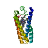 1cpqS S: Starting model for refinement |
|---|---|
| Similar structure data |


-Links
 PDBj
PDBj


- Assembly
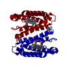
| Deposited unit | 
| ||||||||
|---|---|---|---|---|---|---|---|---|---|
| 1 |
| ||||||||
| Unit cell |
| ||||||||
| Noncrystallographic symmetry (NCS) | NCS oper: (Code: given Matrix: (-0.508819, 0.860871, 0.002075), Vector: |
-Components
| #1: Protein | Mass: 13426.096 Da / Num. of mol.: 2 / Source method: isolated from a natural source / Source: (natural) RHODOBACTER SPHAEROIDES (bacteria) / Cellular location: PERIPLASMIC / References: UniProt: P00148#2: Chemical |   Mass: 618.503 Da / Num. of mol.: 2 / Source method: obtained synthetically / Formula: C34H34FeN4O4 Mass: 618.503 Da / Num. of mol.: 2 / Source method: obtained synthetically / Formula: C34H34FeN4O4#3: Water | ChemComp-HOH / |  Mass: 18.015 Da / Num. of mol.: 291 / Source method: isolated from a natural source / Formula: H2O Mass: 18.015 Da / Num. of mol.: 291 / Source method: isolated from a natural source / Formula: H2OHas protein modification | Y | Sequence details | THERE ARE 2 DIFFERENCES BETWEEN THE DEPOSITORS DATA AND THE GENETIC SEQUENCE(S) : RESIDUE GLY 117 ...THERE ARE 2 DIFFERENCE | |
|---|
-Experimental details
-Experiment
| Experiment | Method: X-RAY DIFFRACTION / Number of used crystals: 1 |
|---|
- Sample preparation
| Crystal | Density Matthews: 2.9 Å3/Da / Density % sol: 57 % | ||||||||||||||||||||||||||||||
|---|---|---|---|---|---|---|---|---|---|---|---|---|---|---|---|---|---|---|---|---|---|---|---|---|---|---|---|---|---|---|---|
| Crystal grow | Method: vapor diffusion, hanging drop / pH: 4.5 Details: 12MG/ML, 5+5 UL HANGING DROP, NA-ACETATE 25MM PH4.5, PEG 8000 15%, pH 4.50 | ||||||||||||||||||||||||||||||
| Crystal grow | *PLUS pH: 4.5 / Method: vapor diffusion, hanging drop | ||||||||||||||||||||||||||||||
| Components of the solutions | *PLUS
|
-Data collection
| Diffraction | Mean temperature: 293 K |
|---|---|
| Diffraction source | Source: ROTATING ANODE / Type: RIGAKU RU200 / Wavelength: 1.54178 |
| Detector | Type: RIGAKU IMAGE PLATE / Detector: IMAGE PLATE / Date: Aug 15, 1999 / Details: COLLIMATOR |
| Radiation | Monochromator: GRAPHITE / Protocol: SINGLE WAVELENGTH / Monochromatic (M) / Laue (L): M / Scattering type: x-ray |
| Radiation wavelength | Wavelength: 1.54178 Å / Relative weight: 1 |
| Reflection | Resolution: 1.8→31 Å / Num. obs: 24077 / % possible obs: 86.5 % / Redundancy: 2.8 % / Rsym value: 0.1 / Net I/σ(I): 8.1 |
| Reflection shell | Resolution: 1.8→1.85 Å / Redundancy: 2.2 % / Mean I/σ(I) obs: 4.6 / Rsym value: 0.16 / % possible all: 56.7 |
| Reflection | *PLUS Highest resolution: 1.8 Å / Lowest resolution: 31 Å / % possible obs: 87 % / Num. measured all: 67416 / Rmerge(I) obs: 0.1 |
| Reflection shell | *PLUS % possible obs: 57 % / Num. unique obs: 2553 / Num. measured obs: 5617 / Rmerge(I) obs: 0.16 / Mean I/σ(I) obs: 4.6 |
- Processing
| Software |
| ||||||||||||||||||||
|---|---|---|---|---|---|---|---|---|---|---|---|---|---|---|---|---|---|---|---|---|---|
| Refinement | Method to determine structure: MOLECULAR REPLACEMENT Starting model: 1CPQ Resolution: 1.8→25 Å / SU B: 5.276 / SU ML: 0.154 / Cross valid method: THROUGHOUT / σ(F): 0 / ESU R: 0.172 / ESU R Free: 0.162 / Details: HYDROGENS HAVE BEEN ADDED IN THE RIDING POSITIONS.
| ||||||||||||||||||||
| Displacement parameters | Biso mean: 24.82 Å2
| ||||||||||||||||||||
| Refinement step | Cycle: LAST / Resolution: 1.8→25 Å
| ||||||||||||||||||||
| Refinement | *PLUS Lowest resolution: 31 Å / % reflection Rfree: 10 % / Rfactor Rfree: 0.254 / Rfactor Rwork: 0.204 | ||||||||||||||||||||
| Solvent computation | *PLUS | ||||||||||||||||||||
| Displacement parameters | *PLUS | ||||||||||||||||||||
| Refine LS restraints | *PLUS
| ||||||||||||||||||||
| LS refinement shell | *PLUS Highest resolution: 1.8 Å / Lowest resolution: 1.85 Å / Rfactor Rfree: 0.351 / Rfactor Rwork: 0.271 |
