 Movie
Movie Controller

[English] 日本語
 Yorodumi
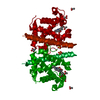
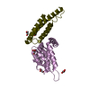
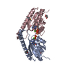
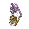
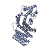
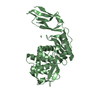
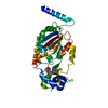
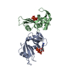
- PDB-1go3: Structure of an archeal homolog of the eukaryotic RNA polymerase ... -
+ Open data

- Basic information
| Entry | Database: PDB / ID: 1go3 | ||||||
|---|---|---|---|---|---|---|---|
| Title | Structure of an archeal homolog of the eukaryotic RNA polymerase II RPB4/RPB7 complex | ||||||
 Components Components |
| ||||||
 Keywords Keywords | TRANSFERASE / TRANSCRIPTION / DNA-DIRECTED RNA POLYMERASE | ||||||
| Function / homology |  Function and homology information Function and homology informationDNA-directed RNA polymerase complex / DNA-templated transcription initiation / DNA-directed RNA polymerase / DNA-directed RNA polymerase activity / nucleotide binding / DNA binding / cytoplasm Similarity search - Function | ||||||
| Biological species |   METHANOCOCCUS JANNASCHII (archaea) METHANOCOCCUS JANNASCHII (archaea) | ||||||
| Method |  X-RAY DIFFRACTION / SYNCHROTRON / MIR / Resolution: 1.75 Å X-RAY DIFFRACTION / SYNCHROTRON / MIR / Resolution: 1.75 Å | ||||||
 Authors Authors | Todone, F. / Brick, P. / Werner, F. / Weinzierl, R.O.J. / Onesti, S. | ||||||
 Citation Citation | Journal: Mol.Cell / Year: 2001 Title: Structure of an Archaeal Homolog of the Eukaryotic RNA Polymerase II Rpb4/Rpb7 Complex Authors: Todone, F. / Brick, P. / Werner, F. / Weinzierl, R.O.J. / Onesti, S. | ||||||
| History |
| ||||||
| Remark 700 | SHEET DETERMINATION METHOD: DSSP THE SHEETS PRESENTED AS "EB" AND "MB" IN EACH CHAIN ON SHEET ... SHEET DETERMINATION METHOD: DSSP THE SHEETS PRESENTED AS "EB" AND "MB" IN EACH CHAIN ON SHEET RECORDS BELOW IS ACTUALLY AN 6-STRANDED BARREL THIS IS REPRESENTED BY A 7-STRANDED SHEET IN WHICH THE FIRST AND LAST STRANDS ARE IDENTICAL. |
- Structure visualization
| Structure viewer | Molecule: MolmilJmol/JSmol |
|---|
- Downloads & links
-Download
| PDBx/mmCIF format | 1go3.cif.gz | 124.2 KB | Display | PDBx/mmCIF format |
|---|---|---|---|---|
| PDB format | pdb1go3.ent.gz | 97.9 KB | Display | PDB format |
| PDBx/mmJSON format | 1go3.json.gz | Tree view | PDBx/mmJSON format | |
| Others |  Other downloads Other downloads |
-Validation report
| Arichive directory | https://data.pdbj.org/pub/pdb/validation_reports/go/1go3ftp://data.pdbj.org/pub/pdb/validation_reports/go/1go3 | HTTPS FTP |
|---|
-Related structure data
| Similar structure data |
|---|


-Links
 PDBj
PDBj


- Assembly
| Deposited unit | 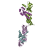
| ||||||||
|---|---|---|---|---|---|---|---|---|---|
| 1 | 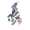
| ||||||||
| 2 | 
| ||||||||
| Unit cell |
| ||||||||
| Noncrystallographic symmetry (NCS) | NCS oper: (Code: given Matrix: (-0.12195, -0.99217, 0.02686), Vector: |
-Components
| #1: Protein | Mass: 21246.625 Da / Num. of mol.: 2 Source method: isolated from a genetically manipulated source Source: (gene. exp.) METHANOCOCCUS JANNASCHII (archaea)Description: CO-EXPRESSED USING A BICISTRIONIC EXPRESSION STRATEGY Plasmid: PGEX-2TK / Production host:  #2: Protein | Mass: 12342.216 Da / Num. of mol.: 2 Source method: isolated from a genetically manipulated source Source: (gene. exp.) METHANOCOCCUS JANNASCHII (archaea)Description: CO-EXPRESSED USING A BICISTRIONIC EXPRESSION STRATEGY Plasmid: PGEX-2TX / Production host: #3: Water | ChemComp-HOH / |  Mass: 18.015 Da / Num. of mol.: 328 / Source method: isolated from a natural source / Formula: H2O Mass: 18.015 Da / Num. of mol.: 328 / Source method: isolated from a natural source / Formula: H2O |
|---|
-Experimental details
-Experiment
| Experiment | Method: X-RAY DIFFRACTION / Number of used crystals: 1 |
|---|
- Sample preparation
| Crystal | Density Matthews: 2.9 Å3/Da / Density % sol: 55 % | |||||||||||||||||||||||||||||||||||||||||||||||||
|---|---|---|---|---|---|---|---|---|---|---|---|---|---|---|---|---|---|---|---|---|---|---|---|---|---|---|---|---|---|---|---|---|---|---|---|---|---|---|---|---|---|---|---|---|---|---|---|---|---|---|
| Crystal grow | pH: 4.6 Details: 100 MM TRIS/ HCL 1.3-1.4 M AMMONIUM DIHYDROGEN MONOPHOSPHATE, pH 4.60 | |||||||||||||||||||||||||||||||||||||||||||||||||
| Crystal grow | *PLUS Temperature: 4 ℃ / pH: 6.5 / Method: vapor diffusion, hanging drop | |||||||||||||||||||||||||||||||||||||||||||||||||
| Components of the solutions | *PLUS
|
-Data collection
| Diffraction | Mean temperature: 100 K |
|---|---|
| Diffraction source | Source: SYNCHROTRON / Site: ESRF  / Beamline: ID14-4 / Wavelength: 1 / Beamline: ID14-4 / Wavelength: 1 |
| Detector | Detector: CCD / Date: Feb 15, 2001 |
| Radiation | Protocol: SINGLE WAVELENGTH / Monochromatic (M) / Laue (L): M / Scattering type: x-ray |
| Radiation wavelength | Wavelength: 1 Å / Relative weight: 1 |
| Reflection | Resolution: 1.75→27.8 Å / Num. obs: 76814 / % possible obs: 99.8 % / Observed criterion σ(I): 3 / Redundancy: 3.6 % / Biso Wilson estimate: 15.4 Å2 / Rmerge(I) obs: 0.092 / Net I/σ(I): 4.1 |
| Reflection shell | Resolution: 1.75→1.85 Å / Redundancy: 3 % / Rmerge(I) obs: 0.234 / Mean I/σ(I) obs: 3 / % possible all: 99.8 |
| Reflection | *PLUS Lowest resolution: 28 Å |
| Reflection shell | *PLUS % possible obs: 99.6 % |
- Processing
| Software |
| ||||||||||||||||||||||||||||||||||||||||||||||||||||||||||||
|---|---|---|---|---|---|---|---|---|---|---|---|---|---|---|---|---|---|---|---|---|---|---|---|---|---|---|---|---|---|---|---|---|---|---|---|---|---|---|---|---|---|---|---|---|---|---|---|---|---|---|---|---|---|---|---|---|---|---|---|---|---|
| Refinement | Method to determine structure: MIR / Resolution: 1.75→27.8 Å / Rfactor Rfree error: 0.004 / Data cutoff high absF: 2534087 / Cross valid method: THROUGHOUT / σ(F): 0
| ||||||||||||||||||||||||||||||||||||||||||||||||||||||||||||
| Solvent computation | Solvent model: FLAT MODE / Bsol: 60.16 Å2 / ksol: 0.39 e/Å3 | ||||||||||||||||||||||||||||||||||||||||||||||||||||||||||||
| Displacement parameters | Biso mean: 20.9 Å2
| ||||||||||||||||||||||||||||||||||||||||||||||||||||||||||||
| Refine analyze |
| ||||||||||||||||||||||||||||||||||||||||||||||||||||||||||||
| Refinement step | Cycle: LAST / Resolution: 1.75→27.8 Å
| ||||||||||||||||||||||||||||||||||||||||||||||||||||||||||||
| Refine LS restraints |
| ||||||||||||||||||||||||||||||||||||||||||||||||||||||||||||
| LS refinement shell | Resolution: 1.75→1.86 Å / Rfactor Rfree error: 0.011 / Total num. of bins used: 6
| ||||||||||||||||||||||||||||||||||||||||||||||||||||||||||||
| Xplor file |
| ||||||||||||||||||||||||||||||||||||||||||||||||||||||||||||
| Software | *PLUS Name: CNS / Version: 1 / Classification: refinement | ||||||||||||||||||||||||||||||||||||||||||||||||||||||||||||
| Refinement | *PLUS Lowest resolution: 20 Å / % reflection Rfree: 5 % | ||||||||||||||||||||||||||||||||||||||||||||||||||||||||||||
| Solvent computation | *PLUS | ||||||||||||||||||||||||||||||||||||||||||||||||||||||||||||
| Displacement parameters | *PLUS | ||||||||||||||||||||||||||||||||||||||||||||||||||||||||||||
| Refine LS restraints | *PLUS
|
