 Movie
Movie Controller

[English] 日本語
 Yorodumi
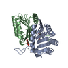
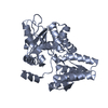
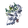
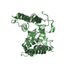
- PDB-1gmz: Crystal structure of the D49 phospholipase A2 piratoxin III from ... -
+ Open data

- Basic information
| Entry | Database: PDB / ID: 1gmz | ||||||
|---|---|---|---|---|---|---|---|
| Title | Crystal structure of the D49 phospholipase A2 piratoxin III from Bothrops pirajai. | ||||||
 Components Components | PHOSPHOLIPASE A2 | ||||||
 Keywords Keywords | HYDROLASE / PHOSPHOLIPASE A2 / NEUROTOXIC / BOTHROPS PIRAJAI | ||||||
| Function / homology |  Function and homology information Function and homology information: / arachidonate secretion / lipid catabolic process / phospholipid metabolic process / phospholipid binding / toxin activity / calcium ion binding / extracellular region Similarity search - Function | ||||||
| Biological species |  BOTHROPS PIRAJAI (snake) BOTHROPS PIRAJAI (snake) | ||||||
| Method |  X-RAY DIFFRACTION / SYNCHROTRON / MOLECULAR REPLACEMENT / Resolution: 2.4 Å X-RAY DIFFRACTION / SYNCHROTRON / MOLECULAR REPLACEMENT / Resolution: 2.4 Å | ||||||
 Authors Authors | Rigden, D.J. / Lee, W.H. / Polikarpov, I. | ||||||
 Citation Citation | Journal: Acta Crystallogr.,Sect.D / Year: 2003 Title: The Structure of the D49 Phospholipase A2 Piratoxin III from Bothrops Pirajai Reveals Unprecedented Structural Displacement of the Calcium-Binding Loop: Possible Relationship to Cooperative Substrate Binding Authors: Rigden, D.J. / Hwa, L. / Marangoni, S. / Toyama, M. / Polikarpov, I. | ||||||
| History |
|
- Structure visualization
| Structure viewer | Molecule: MolmilJmol/JSmol |
|---|
- Downloads & links
-Download
| PDBx/mmCIF format | 1gmz.cif.gz | 62.2 KB | Display | PDBx/mmCIF format |
|---|---|---|---|---|
| PDB format | pdb1gmz.ent.gz | 46.2 KB | Display | PDB format |
| PDBx/mmJSON format | 1gmz.json.gz | Tree view | PDBx/mmJSON format | |
| Others |  Other downloads Other downloads |
-Validation report
| Arichive directory | https://data.pdbj.org/pub/pdb/validation_reports/gm/1gmzftp://data.pdbj.org/pub/pdb/validation_reports/gm/1gmz | HTTPS FTP |
|---|
-Related structure data
| Related structure data | 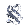 1cl5S S: Starting model for refinement |
|---|---|
| Similar structure data |


-Links
 PDBj
PDBj

- Assembly
| Deposited unit | 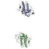
| ||||||||||||
|---|---|---|---|---|---|---|---|---|---|---|---|---|---|
| 1 | 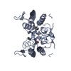
| ||||||||||||
| 2 | 
| ||||||||||||
| Unit cell |
| ||||||||||||
| Components on special symmetry positions |
| ||||||||||||
| Noncrystallographic symmetry (NCS) | NCS oper: (Code: given Matrix: (-0.99992, 0.006062, 0.011136), Vector: |
-Components
| #1: Protein | Mass: 13856.016 Da / Num. of mol.: 2 / Source method: isolated from a natural source / Source: (natural) BOTHROPS PIRAJAI (snake) / Secretion: VENOM / References: UniProt: P58464*PLUS, phospholipase A2#2: Chemical |   Mass: 60.095 Da / Num. of mol.: 2 / Source method: obtained synthetically / Formula: C3H8O / Comment: alkaloid*YM Mass: 60.095 Da / Num. of mol.: 2 / Source method: obtained synthetically / Formula: C3H8O / Comment: alkaloid*YM#3: Water | ChemComp-HOH / |  Mass: 18.015 Da / Num. of mol.: 135 / Source method: isolated from a natural source / Formula: H2O Mass: 18.015 Da / Num. of mol.: 135 / Source method: isolated from a natural source / Formula: H2OHas protein modification | Y | Sequence details | NOT YET DEPOSITED IN SEQUENCE DATABASES RESIDUE UNK(117) IS UNKNOWN IN CHAINS A,B | |
|---|
-Experimental details
-Experiment
| Experiment | Method: X-RAY DIFFRACTION / Number of used crystals: 1 |
|---|
- Sample preparation
| Crystal | Density Matthews: 2.32 Å3/Da / Density % sol: 45 % | |||||||||||||||||||||||||
|---|---|---|---|---|---|---|---|---|---|---|---|---|---|---|---|---|---|---|---|---|---|---|---|---|---|---|
| Crystal grow | pH: 5.3 Details: 18% 2-PROPANOL, 21% PEG 4000 AND 0.1 M NA CITRATE PH 5.3 | |||||||||||||||||||||||||
| Crystal grow | *PLUS Temperature: 291 K / Method: vapor diffusion, hanging drop | |||||||||||||||||||||||||
| Components of the solutions | *PLUS
|
-Data collection
| Diffraction | Mean temperature: 290 K |
|---|---|
| Diffraction source | Source: SYNCHROTRON / Site: LNLS  / Beamline: D03B-MX1 / Wavelength: 1.535 / Beamline: D03B-MX1 / Wavelength: 1.535 |
| Detector | Type: MARRESEARCH / Detector: IMAGE PLATE / Date: Sep 15, 2000 |
| Radiation | Protocol: SINGLE WAVELENGTH / Monochromatic (M) / Laue (L): M / Scattering type: x-ray |
| Radiation wavelength | Wavelength: 1.535 Å / Relative weight: 1 |
| Reflection | Resolution: 2.4→14.7 Å / Num. obs: 8166 / % possible obs: 85.9 % / Redundancy: 2.9 % / Biso Wilson estimate: 20.1 Å2 / Rmerge(I) obs: 0.114 |
| Reflection shell | Resolution: 2.4→2.51 Å / Rmerge(I) obs: 0.264 / % possible all: 89.6 |
| Reflection shell | *PLUS % possible obs: 89.6 % / Redundancy: 2 % / Mean I/σ(I) obs: 2.1 |
- Processing
| Software |
| ||||||||||||||||||||||||||||||||||||||||||||||||||||||||||||||||||||||||||||||||
|---|---|---|---|---|---|---|---|---|---|---|---|---|---|---|---|---|---|---|---|---|---|---|---|---|---|---|---|---|---|---|---|---|---|---|---|---|---|---|---|---|---|---|---|---|---|---|---|---|---|---|---|---|---|---|---|---|---|---|---|---|---|---|---|---|---|---|---|---|---|---|---|---|---|---|---|---|---|---|---|---|---|
| Refinement | Method to determine structure: MOLECULAR REPLACEMENT Starting model: PDB ENTRY 1CL5 Resolution: 2.4→14.66 Å / Rfactor Rfree error: 0.012 / Isotropic thermal model: RESTRAINED / Cross valid method: THROUGHOUT / σ(F): 0
| ||||||||||||||||||||||||||||||||||||||||||||||||||||||||||||||||||||||||||||||||
| Solvent computation | Solvent model: FLAT MODEL / Bsol: 37.6966 Å2 / ksol: 0.321272 e/Å3 | ||||||||||||||||||||||||||||||||||||||||||||||||||||||||||||||||||||||||||||||||
| Displacement parameters | Biso mean: 33.5 Å2
| ||||||||||||||||||||||||||||||||||||||||||||||||||||||||||||||||||||||||||||||||
| Refine analyze |
| ||||||||||||||||||||||||||||||||||||||||||||||||||||||||||||||||||||||||||||||||
| Refinement step | Cycle: LAST / Resolution: 2.4→14.66 Å
| ||||||||||||||||||||||||||||||||||||||||||||||||||||||||||||||||||||||||||||||||
| Refine LS restraints |
| ||||||||||||||||||||||||||||||||||||||||||||||||||||||||||||||||||||||||||||||||
| LS refinement shell | Resolution: 2.4→2.51 Å / Rfactor Rfree error: 0.049 / Total num. of bins used: 8
| ||||||||||||||||||||||||||||||||||||||||||||||||||||||||||||||||||||||||||||||||
| Xplor file |
| ||||||||||||||||||||||||||||||||||||||||||||||||||||||||||||||||||||||||||||||||
| Software | *PLUS Name: CNS / Version: 1 / Classification: refinement | ||||||||||||||||||||||||||||||||||||||||||||||||||||||||||||||||||||||||||||||||
| Refinement | *PLUS σ(F): 0 | ||||||||||||||||||||||||||||||||||||||||||||||||||||||||||||||||||||||||||||||||
| Solvent computation | *PLUS | ||||||||||||||||||||||||||||||||||||||||||||||||||||||||||||||||||||||||||||||||
| Displacement parameters | *PLUS | ||||||||||||||||||||||||||||||||||||||||||||||||||||||||||||||||||||||||||||||||
| Refine LS restraints | *PLUS
|
