 Movie
Movie Controller

[English] 日本語
 Yorodumi
- PDB-1ghd: Crystal structure of the glutaryl-7-aminocephalosporanic acid acy... -
+ Open data

- Basic information
| Entry | Database: PDB / ID: 1ghd | ||||||
|---|---|---|---|---|---|---|---|
| Title | Crystal structure of the glutaryl-7-aminocephalosporanic acid acylase by mad phasing | ||||||
 Components Components | (GLUTARYL-7-AMINOCEPHALOSPORANIC ACID ACYLASE) x 2 | ||||||
 Keywords Keywords | HYDROLASE / cephalosporin acylase | ||||||
| Function / homology |  Function and homology information Function and homology informationhydrolase activity, acting on carbon-nitrogen (but not peptide) bonds, in linear amides / antibiotic biosynthetic process / metal ion binding Similarity search - Function | ||||||
| Biological species |  Pseudomonas sp. 130 (bacteria) Pseudomonas sp. 130 (bacteria) | ||||||
| Method |  X-RAY DIFFRACTION / SYNCHROTRON / Resolution: 2.4 Å X-RAY DIFFRACTION / SYNCHROTRON / Resolution: 2.4 Å | ||||||
 Authors Authors | Ding, Y. / Jiang, W. / Mao, X. / He, H. / Zhang, S. / Tang, H. / Bartlam, M. / Ye, S. / Jiang, F. / Liu, Y. ...Ding, Y. / Jiang, W. / Mao, X. / He, H. / Zhang, S. / Tang, H. / Bartlam, M. / Ye, S. / Jiang, F. / Liu, Y. / Zhao, G. / Rao, Z. | ||||||
 Citation Citation | Journal: J.Biol.Chem. / Year: 2002 Title: Affinity alkylation of the Trp-B4 residue of the beta -subunit of the glutaryl 7-aminocephalosporanic acid acylase of Pseudomonas sp. 130. Authors: Huang, X. / Zeng, R. / Ding, X. / Mao, X. / Ding, Y. / Rao, Z. / Xie, Y. / Jiang, W. / Zhao, G. | ||||||
| History |
|
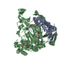
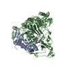
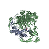
- Structure visualization
| Structure viewer | Molecule: MolmilJmol/JSmol |
|---|
- Downloads & links
-Download
| PDBx/mmCIF format | 1ghd.cif.gz | 146.1 KB | Display | PDBx/mmCIF format |
|---|---|---|---|---|
| PDB format | pdb1ghd.ent.gz | 115.3 KB | Display | PDB format |
| PDBx/mmJSON format | 1ghd.json.gz | Tree view | PDBx/mmJSON format | |
| Others |  Other downloads Other downloads |
-Validation report
| Arichive directory | https://data.pdbj.org/pub/pdb/validation_reports/gh/1ghdftp://data.pdbj.org/pub/pdb/validation_reports/gh/1ghd | HTTPS FTP |
|---|
-Related structure data
| Similar structure data |
|---|


-Links
 PDBj
PDBj


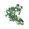
- Assembly
| Deposited unit | 
| ||||||||
|---|---|---|---|---|---|---|---|---|---|
| 1 |
| ||||||||
| 2 | 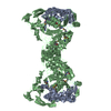
| ||||||||
| Unit cell |
|
-Components
| #1: Protein | Mass: 18737.322 Da / Num. of mol.: 1 / Fragment: ALPHA-SUBUNIT + SPACER PEPTIDE Source method: isolated from a genetically manipulated source Source: (gene. exp.) Pseudomonas sp. 130 (bacteria) / Plasmid: PMFT7H6CAII / Production host: References: UniProt: O86089, Hydrolases; Acting on carbon-nitrogen bonds, other than peptide bonds; In linear amides |
|---|---|
| #2: Protein | Mass: 58855.785 Da / Num. of mol.: 1 / Fragment: BETA-SUBUNIT Source method: isolated from a genetically manipulated source Source: (gene. exp.) Pseudomonas sp. 130 (bacteria) / Plasmid: PMFT7H6CAII / Production host: References: UniProt: O86089, Hydrolases; Acting on carbon-nitrogen bonds, other than peptide bonds; In linear amides |
| #3: Water | ChemComp-HOH /  Mass: 18.015 Da / Num. of mol.: 198 / Source method: isolated from a natural source / Formula: H2O Mass: 18.015 Da / Num. of mol.: 198 / Source method: isolated from a natural source / Formula: H2O |
| Has protein modification | Y |
-Experimental details
-Experiment
| Experiment | Method: X-RAY DIFFRACTION / Number of used crystals: 1 |
|---|
- Sample preparation
| Crystal | Density Matthews: 3.32 Å3/Da / Density % sol: 62.98 % |
|---|---|
| Crystal grow | Temperature: 291 K / Method: hanging drop/vapor diffusion / pH: 7.5 Details: sodium acetate, cadmium sulfate, hepes, pH 7.5, HANGING DROP/VAPOR DIFFUSION, temperature 291.0K |
| Crystal grow | *PLUS Method: unknown / Details: Ichikawa, S., (1981) Agric. Biol. Chem., 45, 2231. |
-Data collection
| Diffraction | Mean temperature: 115 K |
|---|---|
| Diffraction source | Source: SYNCHROTRON / Site: APS  / Beamline: 19-ID / Wavelength: 0.9639 / Beamline: 19-ID / Wavelength: 0.9639 |
| Detector | Type: SBC-2 / Detector: CCD / Date: Nov 8, 2000 |
| Radiation | Protocol: MAD / Monochromatic (M) / Laue (L): M / Scattering type: x-ray |
| Radiation wavelength | Wavelength: 0.9639 Å / Relative weight: 1 |
| Reflection | Resolution: 2.4→100 Å / Num. all: 69082 / Num. obs: 68105 / % possible obs: 98.6 % / Observed criterion σ(I): -3 / Redundancy: 7.6 % / Biso Wilson estimate: 26 Å2 / Rmerge(I) obs: 0.128 / Net I/σ(I): 30.5 |
| Reflection shell | Resolution: 2.4→2.48 Å / Redundancy: 6.2 % / Rmerge(I) obs: 0.48 / Num. unique all: 5097 / % possible all: 97.1 |
- Processing
| Software |
| |||||||||||||||||||||
|---|---|---|---|---|---|---|---|---|---|---|---|---|---|---|---|---|---|---|---|---|---|---|
| Refinement | Resolution: 2.4→30 Å / σ(F): 0 / σ(I): 0 / Stereochemistry target values: Engh & Huber / Details: CNS
| |||||||||||||||||||||
| Refinement step | Cycle: LAST / Resolution: 2.4→30 Å
| |||||||||||||||||||||
| Refine LS restraints |
|
