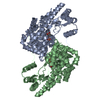
 Movie
Movie Controller


+ Open data

- Basic information
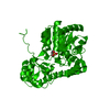
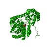
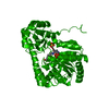
| Entry | Database: PDB / ID: 1g7x | ||||||
|---|---|---|---|---|---|---|---|
| Title | ASPARTATE AMINOTRANSFERASE ACTIVE SITE MUTANT N194A/R292L/R386L | ||||||
 Components Components | ASPARTATE AMINOTRANSFERASE | ||||||
 Keywords Keywords | TRANSFERASE / ACTIVE SITE MUTANT | ||||||
| Function / homology |  Function and homology information Function and homology information: / L-tyrosine:2-oxoglutarate transaminase activity / L-phenylalanine biosynthetic process / aspartate transaminase / L-aspartate:2-oxoglutarate transaminase activity / pyridoxal phosphate binding / protein homodimerization activity / identical protein binding / cytoplasm / cytosol Similarity search - Function | ||||||
| Biological species |  | ||||||
| Method |  X-RAY DIFFRACTION / MOLECULAR REPLACEMENT / Resolution: 2.2 Å X-RAY DIFFRACTION / MOLECULAR REPLACEMENT / Resolution: 2.2 Å | ||||||
 Authors Authors | Mizuguchi, H. / Hayashi, H. / Okada, K. / Miyahara, I. / Hirotsu, K. / Kagamiyama, H. | ||||||
 Citation Citation | Journal: Biochemistry / Year: 2001 Title: Strain is more important than electrostatic interaction in controlling the pKa of the catalytic group in aspartate aminotransferase. Authors: Mizuguchi, H. / Hayashi, H. / Okada, K. / Miyahara, I. / Hirotsu, K. / Kagamiyama, H. | ||||||
| History |
| ||||||
| Remark 999 | SEQUENCE RESIDUE NUMBERS ARE NOT CONTINUOUS THROUGHOUT THE PROTEIN. RESIDUES 64 AND 66, 126 AND ...SEQUENCE RESIDUE NUMBERS ARE NOT CONTINUOUS THROUGHOUT THE PROTEIN. RESIDUES 64 AND 66, 126 AND 129, 152 AND 154, 231 AND 233, & 406 AND 408 ARE COVALENTLY BOUND. |
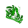
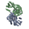
- Structure visualization
| Structure viewer | Molecule: MolmilJmol/JSmol |
|---|
- Downloads & links
-Download
| PDBx/mmCIF format | 1g7x.cif.gz | 94.4 KB | Display | PDBx/mmCIF format |
|---|---|---|---|---|
| PDB format | pdb1g7x.ent.gz | 70 KB | Display | PDB format |
| PDBx/mmJSON format | 1g7x.json.gz | Tree view | PDBx/mmJSON format | |
| Others |  Other downloads Other downloads |
-Validation report
| Arichive directory | https://data.pdbj.org/pub/pdb/validation_reports/g7/1g7xftp://data.pdbj.org/pub/pdb/validation_reports/g7/1g7x | HTTPS FTP |
|---|
-Related structure data
| Related structure data |  1g4vC  1g4xC  1g7wC  1arsS S: Starting model for refinement C: citing same article ( |
|---|---|
| Similar structure data |

-Links
 PDBj
PDBj- Assembly
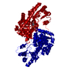
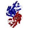
| Deposited unit | 
| ||||||||
|---|---|---|---|---|---|---|---|---|---|
| 1 | 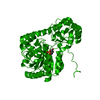
| ||||||||
| Unit cell |
| ||||||||
| Details | The biological assembly is a dimer in the asymmetric unit by the operations: x, -y, -z |
-Components
| #1: Protein | Mass: 43488.109 Da / Num. of mol.: 1 / Mutation: N194A, R292L, R386L Source method: isolated from a genetically manipulated source Source: (gene. exp.) |
|---|---|
| #2: Chemical | ChemComp-PLP /   Mass: 247.142 Da / Num. of mol.: 1 / Source method: obtained synthetically / Formula: C8H10NO6P Mass: 247.142 Da / Num. of mol.: 1 / Source method: obtained synthetically / Formula: C8H10NO6P |
| #3: Water | ChemComp-HOH /  Mass: 18.015 Da / Num. of mol.: 187 / Source method: isolated from a natural source / Formula: H2O Mass: 18.015 Da / Num. of mol.: 187 / Source method: isolated from a natural source / Formula: H2O |
-Experimental details
-Experiment
| Experiment | Method: X-RAY DIFFRACTION / Number of used crystals: 1 |
|---|
- Sample preparation
| Crystal | Density Matthews: 3.19 Å3/Da / Density % sol: 61.39 % | ||||||||||||||||||||||||||||||||||||||||||||||||||||||
|---|---|---|---|---|---|---|---|---|---|---|---|---|---|---|---|---|---|---|---|---|---|---|---|---|---|---|---|---|---|---|---|---|---|---|---|---|---|---|---|---|---|---|---|---|---|---|---|---|---|---|---|---|---|---|---|
| Crystal grow | Temperature: 293 K / Method: vapor diffusion, hanging drop / pH: 7 Details: Sodium Sulfate , pH 7.0, VAPOR DIFFUSION, HANGING DROP, temperature 293K | ||||||||||||||||||||||||||||||||||||||||||||||||||||||
| Crystal grow | *PLUS | ||||||||||||||||||||||||||||||||||||||||||||||||||||||
| Components of the solutions | *PLUS
|
-Data collection
| Diffraction | Mean temperature: 293 K |
|---|---|
| Diffraction source | Source: ROTATING ANODE / Type: RIGAKU RU200 / Wavelength: 1.5418 Å |
| Detector | Type: RIGAKU RAXIS IIC / Detector: IMAGE PLATE / Date: Aug 9, 1995 |
| Radiation | Monochromator: GRAPHITE / Protocol: SINGLE WAVELENGTH / Monochromatic (M) / Laue (L): M / Scattering type: x-ray |
| Radiation wavelength | Wavelength: 1.5418 Å / Relative weight: 1 |
| Reflection | Resolution: 2.2→10 Å / Num. obs: 23701 / % possible obs: 83.8 % / Observed criterion σ(I): 1 / Rmerge(I) obs: 0.069 / Net I/σ(I): 12 |
| Reflection | *PLUS Highest resolution: 2.06 Å / Num. obs: 30457 / % possible obs: 96.2 % / Num. measured all: 101587 / Rmerge(I) obs: 0.087 |
- Processing
| Software |
| ||||||||||||
|---|---|---|---|---|---|---|---|---|---|---|---|---|---|
| Refinement | Method to determine structure: MOLECULAR REPLACEMENT Starting model: PDB ENTRY 1ARS Resolution: 2.2→10 Å / σ(F): 2 / Stereochemistry target values: Engh & Huber /
| ||||||||||||
| Refinement step | Cycle: LAST / Resolution: 2.2→10 Å
| ||||||||||||
| Refine LS restraints |
| ||||||||||||
| Software | *PLUS Name: X-PLOR / Version: 3.1 / Classification: refinement | ||||||||||||
| Refinement | *PLUS Highest resolution: 2.2 Å / Lowest resolution: 10 Å / σ(F): 2 | ||||||||||||
| Solvent computation | *PLUS | ||||||||||||
| Displacement parameters | *PLUS |
